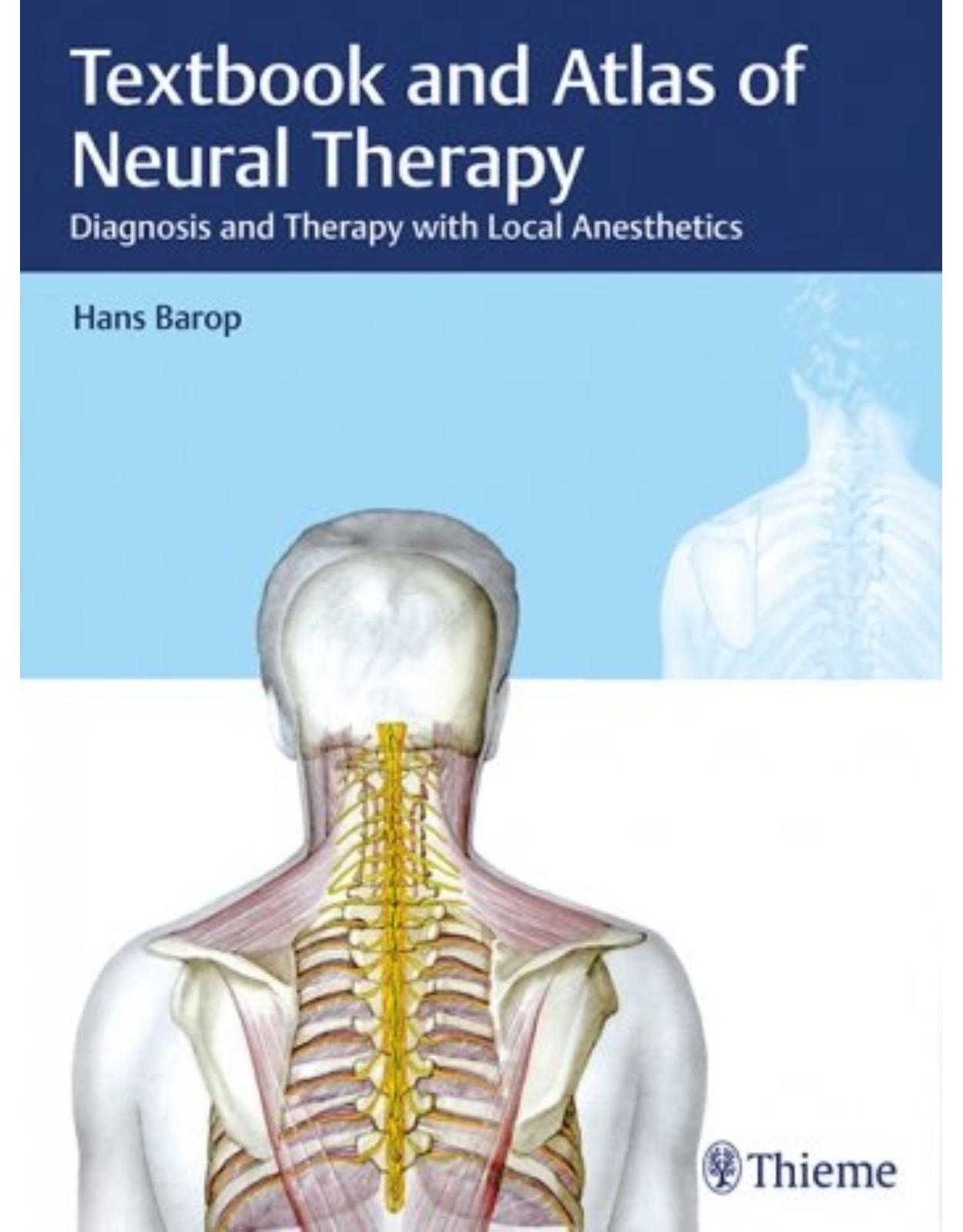
Textbook and Atlas of Neural Therapy: Diagnosis and Therapy with Local Anesthetics
Livrare gratis la comenzi peste 500 RON. Pentru celelalte comenzi livrarea este 20 RON.
Disponibilitate: La comanda in aproximativ 4-6 saptamani
Autor: Hans Barop
Editura: Thieme
Limba: Engleza
Nr. pagini: 343
Coperta: Hardback
Dimensiuni: 27.9 x 21.1 cm
An aparitie: 2017
Description:
Textbook and Atlas of Neural Therapy
Neural therapy is widely used throughout Europe and is rapidly gaining worldwide acceptance in the therapeutic armamentarium. It is known to provide instant relief of pain, improved motion, and return of function and can be used for a variety of health problems unresolved by other methods. This book covers the methodology, indications, and techniques for injecting local anesthetics to specific nerve and tissue sites to restore proper biological function. With this striking visual guide at hand, even physicians without prior experience in neural therapy should be able to implement the techniques successfully in their everyday practice.
Key Features:
Unique "hybrid" illustrations composed of body photographs and drawings of underlying anatomy on the skin clarify the anatomic landmarks for pinpointing the best sites for injections
Useful as a primary teaching tool, but also to add value to the general practitioner's or surgeon's existing practice
Concise explanatory texts on facing pages support the images
Barop's Textbook and Atlas of Neural Therapy will be valued by all students and practitioners involved in the treatment of patients with chronic pain syndromes.
Table of Contents:
Part 1 History and Theory
1 History of Local Anesthesia and Neural Therapy
1.1 Introduction
1.2 Anesthesia and the Treatment of Pain
1.2.1 Local Anesthesia
1.2.2 Neural Therapy
2 Theoretical Foundations and Practice-Based Hypotheses
2.1 Introduction
2.2 Autonomic Nervous System
2.2.1 Anatomy and Function
2.2.2 Sympathetic Efferent
2.2.3 Sympathetic Afferent
2.2.4 Parasympathetic Efferent
2.2.5 Parasympathetic Afferent
2.2.6 Afferent of the Phrenic Nerve
3 Interstitial Regulation System According to Pischinger and Heine
3.1 Introduction
3.2 Structure and Function
3.3 Significance of Autonomic End Formation
3.4 Interstitial Regulation System and Autonomic Nervous System
3.5 Summary
4 Ricker’s Pathology of Relations
4.1 Introduction
4.2 Basics of the Experiments, Stimulus, and Stimulus Effects
4.2.1 Experiments with the Sympathetic Nervous System 18
4.2.2 Particulars of the Sympathetic Nervous System
4.2.3 Medication-Based Stimulus Disruption
4.2.4 Reaction of the Vascular System
4.2.5 Properties of the Blood and its Components
4.2.6 Result of Pathological Sympathetic Nervous System Stimulation
4.2.7 Effect on Specific Tissues
4.3 Ricker’s Three Stage Laws
4.4 Pathology of Relations and Neural Therapy
5 Functional Aspects of the Autonomic Nervous System
5.1 Introduction
5.2 Reaction and Function of the Sympathetic Nervous System
5.3 Therapeutic Use of the Sympathetic Nervous System
6 Concept of the Segment in Neural Therapy
6.1 Definition
6.2 Therapeutic Implications
7 Theory and Basic Principles of the Interference Field
7.1 Introduction
7.1.1 Stimulus and the Sympathetic Nervous System
7.1.2 Causes of Chronic Irritation, Neuroplasticity
7.1.3 Stimulus Interruption—Turning Off the Interference Field
7.2 Pathogenesis of the Interference Field
7.2.1 Temporal Relationships
7.2.2 Emergence of an Interference Field
7.3 Clinical Evidence of the Interference Field
7.4 Interference Field and Segmental Disease
7.4.1 Fluid Transition
7.5 Case Histories and Interpretation
7.6 Neurophysiological Criteria of the Interference Field
7.7 Summary
8 Local Anesthesia in Neural Therapy
8.1 Introduction
8.2 Local Anesthesia as a Neural Therapeutic Agent
8.2.1 Procaine for Neural Therapy
8.2.2 Comparison of Procaine and Lidocaine
8.2.3 The Effects of Procaine Reaction of the Blood Vessels
8.3 Summary
Part 2 The Practice of Neural Therapy
9 Clinical Examination
9.1 Neural Therapeutic Medical History
9.2 Examination
9.2.1 Skin
9.2.2 Musculoskeletal System
9.2.3 Oral Cavity and Teeth
9.3 Palpation
9.4 Other Examination Options
9.5 Documentation
9.6 Structure of Neural Therapeutic Practice
9.7 Selecting the Neural Therapeutic Agent
10 Segments
10.1 Segment Diagnostics—Segmental Therapy
10.2 Lung Segment
10.2.1 Diagnostics
10.2.2 Therapy
10.2.3 Summary
10.3 Heart Segment
10.3.1 Diagnostics
10.3.2 Therapy
10.3.3 Summary
10.4 Hepatobiliary Segment
10.4.1 Diagnostics
10.4.2 Therapy
10.4.3 Summary
10.5 Stomach Segment
10.5.1 Diagnostics
10.5.2 Therapy
10.5.3 Summary
10.6 Pancreatic Segment
10.6.1 Diagnostics
10.6.2 Therapy
10.6.3 Summary
10.7 Intestinal Segment
10.7.1 Diagnostics
10.7.2 Therapy
10.7.3 Summary
10.8 Kidney Segment
10.8.1 Diagnostics
10.8.2 Therapy
10.8.3 Summary
11 Segment Diagnostics
11.1 Tabular Overview
12 Interference Field
12.1 Interference Field Diagnostics
12.2 Classification
12.3 Interference Field Therapy
12.4 Basic Principles
12.5 The Most Common Interference Fields
13 Maxillodental Region
13.1 Example 1
13.2 Example 2
13.3 Example 3
13.4 Example 4
13.5 Example 5
13.6 Example 6
13.7 Summary
14 Neural Therapeutic Phenomena
14.1 Neural Therapeutic Phenomena and Types of Reactions
14.1.1 Segment Phenomenon
14.1.2 Reaction Phenomenon (According to Hopfer)
14.1.3 Retrograde Phenomenon (According to Hopfer)
14.1.4 Second Phenomenon (Huneke’s Phenomenon)
14.1.5 Delayed Second Phenomenon
14.1.6 Incomplete Second Phenomenon
14.1.7 “Silent” Second Phenomenon
14.2 Tactical Approach
14.3 Side Effects
14.4 Failure of Neural Therapy
14.4.1 Causes
14.4.2 Further Diagnostic and Therapeutic Possibilities
Part 3 Injection Technique and Indications
15 General Information
15.1 Introduction
15.2 Positioning of the Patient
15.3 Disinfection
15.4 Injection Method
15.5 Briefing
15.6 Complications, Risks, and Errors
15.7 Dosage of the Local Anesthetic
15.8 Frequent Injections
15.8.1 The Wheal
15.8.2 Infiltration of Geloses
15.8.3 Injection in Muscular Trigger Points and Muscle Insertions
15.8.4 Infiltration of Trigger Points
15.8.5 Infiltration of Scars
15.8.6 The Intravenous Injection
16 Head
16.1 Injection under the Scalp
16.1.1 Indications
16.1.2 Anatomy and Neurophysiology
16.1.3 Injection Technique
16.1.4 Material
16.2 Injection to the Branches of the Trigeminal Nerve 112
16.2.1 Indications
16.2.2 Anatomy and Neurophysiology
16.2.3 Injection Technique
16.2.4 Complications
16.2.5 Material
16.3 Injection to the Mastoid
16.3.1 Indications
16.3.2 Anatomy and Neurophysiology
16.3.3 Injection Technique
16.3.4 Material
16.4 Injection to the Facial Artery, Superficial Temporal Artery, and Auriculotemporal Nerve (from the Mandibular Nerve/Trigeminal Nerve)
16.4.1 Indications
16.4.2 Anatomy and Neurophysiology
16.4.3 Injection Technique
16.4.4 Material
16.5 Injection at and in the Parotid Gland
16.5.1 Indications
16.5.2 Anatomy and Neurophysiology
16.5.3 Injection Technique
16.5.4 Material
16.6 Injection at the Jaw Joint
16.6.1 Indications
16.6.2 Anatomy and Neurophysiology
16.6.3 Injection Technique
16.6.4 Material
16.7 Injection to the Ciliary Ganglion (Retrobulbar Injection)
16.7.1 Indications
16.7.2 Anatomy and Neurophysiology
16.7.3 Injection Technique
16.7.4 Complications
16.7.5 Material
16.8 Injection to the Pterygopalatine Ganglion, the Maxillary Nerve, and Maxillary Artery
16.8.1 Indications
16.8.2 Anatomy and Neurophysiology
16.8.3 Injection Technique
16.8.4 Complications
16.8.5 Material
16.9 Injection to the Otic Ganglion and the Mandibular Nerve
16.9.1 Indications
16.9.2 Anatomy and Neurophysiology
16.9.3 Injection Technique (According to Hauberrisser)
16.9.4 Material
16.10 Injection to the Major Occipital Nerve, Occipital Artery, and Minor Occipital Nerve
16.10.1 Indications
16.10.2 Anatomy and Neurophysiology
16.10.3 Injection Technique
16.10.4 Material
16.11 Injection into the Lymphatic Drainage Area of the Facial Skull
16.11.1 Indications
16.11.2 Anatomy and Neurophysiology
16.11.3 Injection Technique
16.11.4 Material
16.12 Injection to the Tonsils
16.12.1 Indications
16.12.2 Anatomy and Neurophysiology
16.12.3 Injection Technique
16.12.4 Material
16.13 Injection to the Teeth
16.13.1 Indications
16.13.2 Anatomy and Neurophysiology
16.13.3 Injection Technique
16.13.4 Material
17 Neck
17.1 Injection in the Thyroid Gland
17.1.1 Indications
17.1.2 Anatomy and Neurophysiology
17.1.3 Injection Technique
17.1.4 Material
17.2 Injection to the Superior Laryngeal Nerve
17.2.1 Indications
17.2.2 Anatomy and Neurophysiology
17.2.3 Injection Technique
17.2.4 Material
17.3 Injection to the Stellate Ganglion (Cervical Thoracic Ganglion)
17.3.1 Indications
17.3.2 Anatomy and Neurophysiology
17.3.3 Injection Technique
17.3.4 Material
17.4 Injection to the Superior Cervical Ganglion
17.4.1 Indications
17.4.2 Anatomy and Neurophysiology
17.4.3 Injection Technique
17.4.4 Material
17.5 Injection to the Accessory Nerve, Great Auricular Nerve, Transverse Cervical Nerve, and Lesser Occipital Nerve (Punctum Nervosum)
17.5.1 Indications
17.5.2 Anatomy and Neurophysiology
17.5.3 Injection Technique
17.5.4 Material
18 Spine
18.1 Information on Diagnostics
18.2 Information on Therapy
18.3 Injection to the Cervical Spine
18.3.1 Indications
18.3.2 Anatomy and Neurophysiology
18.3.3 Injection Technique
18.3.4 Material
18.4 Injection to the Thoracic Spine
18.4.1 Indications
18.4.2 Anatomy and Neurophysiology
18.4.3 Injection Technique
18.4.4 Material
18.5 Injection to the Lumbar Spine
18.5.1 Indications
18.5.2 Anatomy and Neurophysiology
18.5.3 Injection Technique
18.5.4 Material
18.6 Injection to the Spinal Roots L1–S3 (Lumbosacral Plexus)
18.6.1 Introduction
18.6.2 Injection to the Spinal Roots L1–L4 (Lumbar Plexus)
18.6.3 Material
18.6.4 Injection to the Spinal Roots L5–S3 (Sacral Plexus)
18.7 Injections in the Pelvic Region
18.7.1 Indications
18.7.2 Anatomy and Neurophysiology
18.7.3 Injection Technique
18.7.4 Material
18.8 Injection to the Lumbar Sympathetic Trunk
18.8.1 Indications
18.8.2 Anatomy and Neurophysiology
18.8.3 Injection Technique
18.8.4 Material
18.9 Injection in the Sacral and Lumbar Epidural Space
18.9.1 Indications
18.9.2 Anatomy and Neurophysiology
18.9.3 Injection Technique
18.9.4 Material
19 Abdomen, Retroperitoneum
19.1 Injection to the Renal Hilum and the Renal Plexus
19.1.1 Indications
19.1.2 Anatomy and Neurophysiology
19.1.3 Injection Technique
19.1.4 Material
19.2 Injection to the Celiac Ganglion and the Major and Minor Splanchnic Nerves
19.2.1 Indications
19.2.2 Anatomy and Neurophysiology
19.2.3 Injection Technique
19.2.4 Material
19.3 Injection to the Branches of the Inferior Hypogastric Plexus (Pelvic Plexus)
19.3.1 Indications
19.3.2 Anatomy and Neurophysiology
19.3.3 Injection Technique
19.3.4 Complications
19.3.5 Material
19.4 Injection to the Branches of the Prostatic Plexus, in the Prostate
19.4.1 Indications
19.4.2 Anatomy and Neurophysiology
19.4.3 Injection Technique
19.4.4 Complications
19.4.5 Material
20 Joints
20.1 Injection to the Shoulder Joint and Shoulder Girdle
20.1.1 Indications
20.1.2 Anatomy and Neurophysiology
20.1.3 Injection Technique
20.1.4 Material
20.2 Injection to the Elbow Joint
20.2.1 Indications
20.2.2 Anatomy and Neurophysiology
20.2.3 Injection Technique
20.2.4 Material
20.3 Injection to the Wrist and to the Finger Joints
20.3.1 Indications
20.3.2 Anatomy and Neurophysiology
20.3.3 Injection Technique
20.3.4 Material
20.4 Injection to the Hip Joint
20.4.1 Indications
20.4.2 Anatomy and Neurophysiology
20.4.3 Injection Technique
20.4.4 Material
20.5 Injection to the Knee Joint
20.5.1 Indications
20.5.2 Anatomy and Neurophysiology
20.5.3 Injection Technique
20.5.4 Material
20.6 Injection to the Upper and Lower Ankle Joints, the Tarsal/Metatarsal Joints, and the Toe Joints
20.6.1 Indications
20.6.2 Anatomy and Neurophysiology
20.6.3 Injection Technique
20.6.4 Material
Part 4 Indications and Therapy
21 Indications and Therapy
21.1 Introduction
22 Head
22.1 Headache
22.1.1 Diagnoses
22.1.2 Therapy
22.2 Neuralgias
22.2.1 Diagnoses
22.2.2 Therapy
22.3 Illnesses and Injuries of the Brain
22.3.1 Diagnoses
22.3.2 Therapy
22.4 Eye Disorders
22.4.1 Diagnoses
22.4.2 Therapy
22.5 Disorders of the Nose and Paranasal Sinuses
22.5.1 Diagnoses
22.5.2 Therapy
22.6 Disorders of the Ear and Vestibular Organ
22.6.1 Diagnoses
22.6.2 Therapy
22.7 Disorders of the Mouth and Throat
22.7.1 Tonsils and Pharynx
22.7.2 Salivary Glands, Oral, and Pharyngeal Mucosa
22.7.3 Teeth, Periodontal Apparatus, and Gums
23 Neck
23.1 Disorders and Dysfunctions of the Thyroid
23.1.1 Diagnoses
23.1.2 Therapy
23.2 Disorders and Dysfunctions of the Larynx
23.2.1 Diagnoses
23.2.2 Therapy
24 Thorax
24.1 Disorders of the Bronchia and Lungs
24.1.1 Diagnoses
24.1.2 Therapy
24.2 Disorders of the Heart and the Mediastinal Space
24.2.1 Diagnoses
24.2.2 Therapy
24.3 Mammary Gland Disorders
24.3.1 Diagnoses
24.3.2 Therapy
25 Abdomen, Small Pelvis, and Retroperitoneum
25.1 Stomach Disorders
25.1.1 Diagnoses
25.1.2 Therapy
25.2 Disorders of the Small and Large Intestine
25.2.1 Diagnoses
25.2.2 Therapy
25.3 Disorders of the Liver and Bile Ducts
25.3.1 Diagnoses
25.3.2 Therapy
25.4 Disorders of the Pancreas
25.4.1 Diagnoses
25.4.2 Therapy
25.5 Disorders of the Kidneys and Efferent Urinary Tracts
25.5.1 Diagnoses
25.5.2 Therapy
25.6 Disorders of the Female Internal Genitals
25.6.1 Diagnoses
25.6.2 Therapy
25.7 Disorders of the Male Internal and External Genitals
25.7.1 Diagnoses
25.7.2 Therapy
26 Spine and Pelvis
26.1 Degenerative and Inflammatory Diseases, Injuries
26.1.1 Diagnoses
26.1.2 Therapy
27 Extremities and Joints
27.1 Degenerative Disorders, Inflammations, and Injuries
27.1.1 Diagnoses
27.1.2 Therapy
28 Nerves
28.1 Disorders of the Peripheral Nerves and Cranial Nerves
28.1.1 Diagnoses
28.1.2 Therapy
29 Vessels
29.1 Disorders of the Arterial Vessels
29.1.1 Diagnoses
29.1.2 Therapy
29.2 Disorders of the Venous Vessels
29.2.1 Diagnoses
29.2.2 Therapy
30 Lymphatic System
30.1 Disorders of the Lymphatic Channels and Lymph Nodes
30.1.1 Diagnoses
30.1.2 Therapy
31 Skin
31.1 Disorders and Injuries of the Skin and Its Appendages
31.1.1 Diagnoses
31.1.2 Therapy
32 Tumors
32.1 Malignant Diseases
33 Summary
Part 5 Appendix
34 Literature
Index
| An aparitie | 2017 |
| Autor | Hans Barop |
| Dimensiuni | 27.9 x 21.1 cm |
| Editura | Thieme |
| Format | Hardback |
| ISBN | 9783132410497 |
| Limba | Engleza |
| Nr pag | 343 |
-
38600 lei 34900 lei
-
27400 lei 22200 lei








Clientii ebookshop.ro nu au adaugat inca opinii pentru acest produs. Fii primul care adauga o parere, folosind formularul de mai jos.