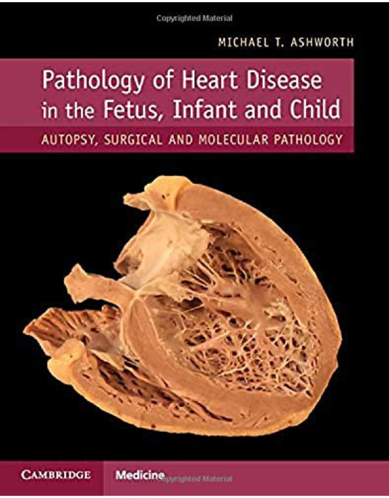
Pathology of Heart Disease in the Fetus, Infant and Child
Livrare gratis la comenzi peste 500 RON. Pentru celelalte comenzi livrarea este 20 RON.
Disponibilitate: La comanda in aproximativ 4-6 saptamani
Autor: Michael T. Ashworth
Editura: Cambridge Unversity Press
Limba: Engleza
Nr. pagini: 500
Coperta: Hardcover
Dimensiuni: 22.7 x 2.2 x 28.3 cm
An aparitie: 22 Aug. 2019
Description:
Uniquely addresses the long overdue need for an authoritative, immersive approach to paediatric cardiac pathology. For general and paediatric pathologists, this book covers all aspects of diseases and malformations of the heart. Extensively illustrated, it approaches work from the fetus to the adolescent, including the effects of surgery.
1. Table of contents:
2. Preface
3. Chapter 1 The Anatomy of the Normal Heart
4. 1.1 Introduction
5. 1.2 Anatomy
6. 1.2.1 Situation
7. 1.2.2 Pericardium
8. 1.2.3 The Right and Left Atrium
9. 1.2.4 The Ventricles
10. 1.2.5 Atrioventricular Valves
11. 1.2.6 Interventricular Septum
12. 1.2.7 Cardiac Conduction System
13. 1.2.8 Arterial Valves
14. 1.2.9 Coronary Arteries
15. 1.2.10 Cardiac Veins
16. 1.3 Histology
17. 1.3.1 Pericardium
18. 1.3.2 Myocardium
19. 1.3.3 Endocardium
20. 1.3.4 Valves
21. 1.3.5 Conduction Tissue
22. 1.3.6 Coronary Arteries
23. 1.3.7 Cardiac Veins
24. 1.3.8 Cardiac Lymphatics
25. 1.3.9 Aorta, Pulmonary Arteries and Arterial Duct
26. 1.3.10 Nerves
27. 1.4 Electron Microscopy
28. 1.5 Weights and Measures
29. References
30. Chapter 2 Examination of the Heart
31. 2.1 Introduction
32. 2.2 Dissection
33. 2.3 Sequential Segmental Analysis
34. 2.3.1 Situs
35. 2.3.2 Topology
36. 2.3.3 Segmental Connections
37. 2.3.4 Atrioventricular Connections
38. 2.3.5 Ventriculoarterial Connections
39. 2.4 Simulated Echocardiographic Views
40. 2.4.1 Another Method for Opening and Sampling the Post-Mortem Heart
41. 2.5 Histology
42. 2.5.1 Sampling for Histology
43. 2.5.2 Endomyocardial Biopsy
44. 2.5.3 Myectomy
45. 2.5.4 Apical Biopsy
46. 2.5.5 Cardiac Tumours
47. 2.5.6 Atrial Appendages
48. 2.5.7 Aorta
49. 2.5.8 Vascular Grafts
50. 2.5.9 Native Valves
51. 2.6 Photography
52. References
53. Chapter 3 Development of the Heart
54. 3.1 Introduction
55. 3.2 Brief Recap of Relevant Early Human Embryonic Development
56. 3.3 Brief Summary of Heart Development
57. 3.4 Early Development
58. 3.4.1 The Heart Fields
59. 3.4.2 The Heart Tube
60. 3.4.3 Contraction
61. 3.5 Looping of the Heart Tube
62. 3.6 Development of the Chambers and Septation
63. 3.6.1 Atrial Septation
64. 3.6.2 The Interventricular Septum
65. 3.6.3 The Atrioventricular Junction
66. 3.6.4 Outflow Tract
67. 3.7 Pericardium
68. 3.8 Coronary Arteries
69. 3.9 Conduction Tissue
70. 3.10 Arterial System
71. 3.11 Venous System
72. 3.11.1 Vitelline Veins
73. 3.11.2 Umbilical Veins
74. 3.11.3 Cardinal Veins
75. 3.12 The Fetal Circulation and Changes at Birth
76. 3.12.1 The Venous Duct and Oval Foramen
77. 3.12.2 Arterial Duct, Lungs and Systemic Circulation
78. 3.12.3 Post-Natal Adaptation
79. References
80. Chapter 4 Congenital Heart Disease (I)
81. 4.1 Introduction
82. 4.2 Ventricular Septal Defect (VSD)
83. 4.3 Atrioventricular Septal Defect (AVSD)
84. 4.4 Atrial Septal Defect (ASD)
85. 4.5 Abnormalities of the Arterial Duct
86. 4.5.1 Absence of the Arterial Duct
87. 4.5.2 Normal Closure
88. 4.5.3 Premature Closure in Utero
89. 4.5.4 Persistent Patency of the Duct
90. 4.5.5 Aneurysm of the Duct
91. 4.6 Coarctation of the Aorta
92. 4.7 Pulmonary Stenosis and Atresia, Including Tetralogy of Fallot
93. 4.7.1 Pulmonary Atresia with Intact Interventricular Septum
94. 4.7.2 Pulmonary Stenosis with Intact Interventricular Septum
95. 4.7.3 Pulmonary Stenosis with VSD, Including Tetralogy of Fallot
96. 4.7.4 Pulmonary Atresia with Ventricular Septal Defect
97. 4.7.5 Absence of the Pulmonary Valve
98. 4.8 Aortic Stenosis
99. 4.8.1 Valvar Stenosis
100. 4.8.2 Subvalvar Stenosis
101. 4.8.3 Supravalvar Stenosis
102. 4.9 Hypoplastic Left Heart
103. 4.10 Transposition of the Great Arteries
104. 4.10.1 Complete Transposition
105. 4.10.2 Arterial Switch Operation
106. 4.10.3 Atrial Switch Operations (Mustard and Senning)
107. 4.10.4 Rastelli Operation
108. 4.10.5 Congenitally Corrected Transposition
109. 4.11 Common Arterial Trunk (Truncus Arteriosus)
110. References
111. Chapter 5 Congenital Heart Disease (II)
112. 5.1 Double Inlet Ventricle
113. 5.1.1 Double Inlet Left Ventricle
114. 5.1.2 Double Inlet Right Ventricle
115. 5.2 Double Outlet Ventricle
116. 5.2.1 Double Outlet Right Ventricle (DORV)
117. 5.3 Abnormalities of the Pulmonary Veins
118. 5.3.1 A Preliminary Note on Terminology
119. 5.3.2 Anomalous Pulmonary Venous Connection
120. 5.3.3 Pulmonary Vein Stenosis
121. 5.4 Ebstein's Malformation
122. 5.5 Tricuspid Atresia
123. 5.6 Other Abnormalities of the Tricuspid Valve
124. 5.6.1 Unguarded Tricuspid Orifice
125. 5.6.2 Absent Commissure
126. 5.7 Uhl's Anomaly
127. 5.8 Atrial Isomerism
128. 5.8.1 Right Atrial Isomerism
129. 5.8.2 Left Atrial Isomerism
130. 5.8.3 Juxtaposition of the Atrial Appendages
131. 5.9 Structural Abnormalities of the Coronary Arteries
132. 5.10 Other Abnormalities
133. 5.10.1 Persistent Left Superior Caval Vein
134. 5.10.2 Aberrant Origin of Right Subclavian Artery
135. 5.10.3 Kommerell's Diverticulum
136. 5.10.4 Ectopia Cordis
137. 5.10.5 Left Atrial Aneurysm
138. 5.11 Anomalies of the Venous Duct (Ductus Venosus)
139. 5.11.1 Absence of the Venous Duct
140. 5.11.2 Persistent Patency of the Venous Duct
141. 5.11.3 Aneurysm of the Sinus of Valsalva
142. 5.11.4 Aorto-Left Ventricular Tunnel
143. 5.11.5 Cor Triatriatum
144. 5.11.6 Aneurysm of Left Ventricle and Left Ventricular Diverticulum
145. 5.12 Pulmonary Vascular Disease in Congenital Heart Disease
146. 5.12.1 Normal Histology
147. 5.12.2 Histopathological Features of Pulmonary Hypertension
148. 5.12.3 Congenital Heart Disease and Pulmonary Vascular Disease
149. 5.12.4 Pulmonary Venous Hypertension
150. 5.12.4.1 Pulmonary Vein Stenosis
151. 5.12.4.2 Obstructed Total Anomalous Pulmonary Venous Connection
152. 5.13 Surgical Operations for Congenital Heart Disease
153. 5.13.1 Patches
154. 5.13.2 Modified Blalock-Taussig Shunt
155. 5.13.3 Bidirectional Glenn Shunt
156. 5.13.4 Damus–Kaye–Stansel (DKS) Procedure
157. 5.13.5 Norwood Procedure
158. 5.13.6 Fontan Procedure
159. 5.13.7 Ross–Konno Procedure
160. 5.13.8 Arterial Switch Operation
161. 5.13.9 Rastelli Operation
162. 5.13.10 Mustard and Senning Operations
163. 5.13.10.1 Mustard Operation
164. 5.13.10.2 Senning Operation
165. 5.13.11 Pulmonary Artery Band
166. 5.13.12 Prosthetic Heart Valves
167. 5.14 Assessment of the Operated Heart
168. References
169. Chapter 6 Ischaemia and Infarction
170. 6.1 Introduction
171. 6.2 Macroscopic Appearance
172. 6.2.1 Subendocardial Necrosis
173. 6.2.2 Papillary Muscle Rupture
174. 6.2.3 Regional Infarction
175. 6.2.4 Coronary Artery Atherosclerosis
176. 6.3 Microscopic Appearance
177. 6.3.1 Dating of Injury
178. 6.3.2 Haemolytic Uraemic Syndrome
179. 6.3.3 Antiphospholipid Syndrome
180. 6.3.4 Myocardial Calcification
181. References
182. Chapter 7 Cardiomyopathy
183. 7.1 Introduction
184. 7.2 Hypertrophic Cardiomyopathy
185. 7.3 Other Cardiomyopathies with a Hypertrophic Phenotype
186. 7.3.1 Friedreich's Ataxia
187. 7.3.2 Noonan's Syndrome
188. 7.4 Dilated Cardiomyopathy
189. 7.5 Restrictive Cardiomyopathy
190. 7.6 Eosinophilic Endomyocardial Disease
191. 7.7 Mitochondrial Cardiomyopathy
192. 7.8 Arrhythmogenic Cardiomyopathy
193. 7.9 Non-Compaction of the Ventricular Myocardium
194. 7.10 Histiocytoid Cardiomyopathy
195. 7.11 Other Forms of Cardiomyopathy
196. References
197. Chapter 8 Inflammation of the Myocardium, Endocardium and Aorta
198. 8.1 Introduction
199. 8.2 Myocarditis
200. 8.2.1 Macroscopic Pathology
201. 8.2.2 Microscopic Pathology
202. 8.2.3 The Dallas Criteria
203. 8.2.4 Giant Cell Myocarditis
204. 8.2.5 Eosinophilic Myocarditis
205. 8.2.6 Bacterial and Protozoal Myocarditis
206. 8.2.6.1 Bacterial Myocarditis
207. 8.2.6.2 Toxoplasma
208. 8.2.6.3 Chagas Disease
209. 8.3 Systemic Inflammatory Diseases with Heart Involvement
210. 8.3.1 Rheumatic Disease
211. 8.3.2 Lupus Erythematosus
212. 8.3.3 Systemic Sclerosis
213. 8.3.4 Juvenile Idiopathic Arthritis
214. 8.3.5 Sarcoidosis
215. 8.4 Aortitis
216. 8.4.1 Takayasu Arteritis
217. 8.4.2 Infectious Aortitis
218. 8.5 Endocarditis
219. 8.5.1 Infectious Endocarditis
220. 8.5.2 Thrombotic Endocarditis
221. References
222. Chapter 9 The Coronary Arteries
223. 9.1 Introduction
224. 9.2 Normal Structure
225. 9.3 Common Normal Variants of the Coronary Arteries
226. 9.4 Abnormal Variations in the Epicardial Distribution of the Coronary Arteries in the Normally Form
227. 9.4.1 Anomalous Origin of the Coronary Arteries from the Pulmonary Artery
228. 9.4.2 Pathological Anomalous Origin of the Coronary Arteries from the Aorta
229. 9.5 Coronary Artery Fistula
230. 9.6 Coronary Artery Hypoplasia and Atresia
231. 9.7 Variations in the Epicardial Coronary Arteries in Congenital Heart Disease
232. 9.7.1 Tetralogy of Fallot
233. 9.7.2 Transposition of the Great Arteries
234. 9.7.3 Common Arterial Trunk
235. 9.7.4 Double Outlet Right Ventricle (DORV)
236. 9.7.5 Hypoplastic Left Heart
237. 9.7.6 Congenitally Corrected Transposition
238. 9.8 Vasculitis Including Kawasaki Disease
239. 9.8.1 Kawasaki Disease
240. 9.8.2 Polyarteritis Nodosa
241. 9.9 Eosinophilic Granulomatosis with Polyangiitis (Formerly Churg-Strauss Syndrome)
242. 9.10 Thrombosis and Embolism
243. 9.11 Fibromuscular Dysplasia
244. 9.12 Segmental Arterial Mediolysis
245. 9.13 Idiopathic Arterial Calcification
246. References
247. Chapter 10 Metabolic and Storage Disease
248. 10.1 Introduction
249. 10.2 Glycogen Storage Disorders
250. 10.2.1 Brief Overview of Glycogen Metabolism (Figure 10.1)
251. 10.2.2 Pompe Disease (Glycogen Storage Disease (GSD) II)
252. 10.2.3 Danon Disease
253. 10.2.4 GSD III (Cori Disease, Debranching Enzyme)
254. 10.2.5 GSD IV (Andersen Disease, Branching Enzyme)
255. 10.2.6 GSD IX (Phosphorylase b Kinase Deficiency)
256. 10.2.7 GSD XV (Glycogenin Deficiency)
257. 10.2.8 GSD 0 (Glycogen Synthase Deficiency)
258. 10.2.9 PRKAG2 Deficiency
259. 10.2.10 Polyglucosan Storage Disease
260. 10.3 Lysosomal Storage Disorders
261. 10.3.1 Anderson–Fabry Disease
262. 10.3.2 Gaucher Disease
263. 10.3.3 Niemann–Pick Disease
264. 10.4 Mucopolysaccharidosis
265. 10.5 Disorders of Fatty Acid Metabolism
266. 10.5.1 Medium-Chain Acyl Co-A Dehydrogenase Deficiency (MCAD)
267. 10.5.2 Very-Long-Chain Acyl Co-A Dehydrogenase Deficiency (VLCHAD)
268. 10.5.3 Trifunctional Protein Deficiency and Long-Chain 3-hydroxyacyl Co-A Dehydrogenase Deficiency (
269. 10.5.4 Carnitine Deficiency
270. 10.5.5 Carnitine-Acylcarnitine Translocase Deficiency
271. 10.5.6 Carnitine Palmitoyl Transferase 2 Deficiency
272. 10.6 Congenital Disorders of Glycosylation
273. 10.7 Disorders of Iron Metabolism
274. 10.7.1 Hereditary Haemochromatosis
275. 10.7.2 Neonatal Haemochromatosis/Gestational Alloimmune Liver Disease
276. 10.8 Organic Acidaemias and Disorders of Amino Acid Metabolism
277. 10.8.1 Propionic Acidaemia
278. 10.8.2 Methylmalonic Aciduria
279. 10.8.3 Methylglutaconic Aciduria
280. 10.8.4 Tyrosinaemia
281. 10.8.5 Oxalosis
282. 10.8.6 Homocystinuria
283. References
284. Chapter 11 Pericardium
285. 11.1 Introduction
286. 11.2 Congenital Defects of the Pericardium
287. 11.3 Cysts and Diverticula
288. 11.4 Heterotopia
289. 11.5 Effusions and Tamponade
290. 11.6 Epicardial Haemorrhage
291. 11.7 Haemopericardium
292. 11.8 Pneumopericardium
293. 11.9 Pericarditis
294. 11.9.1 Acute Purulent Pericarditis
295. 11.9.2 Tuberculous Pericarditis
296. 11.9.3 Other Causes of Pericarditis
297. 11.9.4 Uraemic Pericarditis
298. 11.10 Post-pericardiotomy Syndrome
299. 11.11 Constrictive Pericarditis
300. 11.12 Pericardial Tumours
301. References
302. Chapter 12 Fetal Cardiovascular Disease
303. 12.1 Introduction
304. 12.2 The Normal Fetal Heart
305. 12.3 Fetal Hydrops
306. 12.3.1 Premature Shunt Closure
307. 12.3.2 Arrhythmia
308. 12.3.3 Maternal Lupus
309. 12.4 Syndromes with Heart Malformations
310. 12.4.1 Chromosomal Abnormality
311. 12.4.1.1 Down's Syndrome: Trisomy 21
312. 12.4.1.2 Trisomy 18
313. 12.4.1.3 Trisomy 13
314. 12.4.1.4 Triploidy
315. 12.4.1.5 Turner's Syndrome (45,XO)
316. 12.4.1.6 Other Chromosomal Abnormalities
317. 12.4.1.7 22q11.2 Microdeletion Syndrome
318. 12.4.1.8 Arteriohepatic Dysplasia (Alagille Syndrome)
319. 12.4.1.9 CHARGE Syndrome
320. 12.4.1.10 Noonan's Syndrome
321. 12.4.1.11 Williams Syndrome
322. 12.4.1.12 Marfan Syndrome
323. 12.4.1.13 VACTERL/VATER Association
324. 12.4.1.14 Holt-Oram Syndrome
325. 12.4.1.15 Carney Complex
326. 12.4.1.16 Loeys-Dietz Syndrome
327. 12.4.1.17 Ehlers-Danlos Syndrome
328. 12.4.1.18 Homozygous Familial Hypercholesterolaemia
329. 12.5 Structural Heart Disease in the Fetus
330. 12.6 Fetal Cardiomyopathy
331. 12.7 Fetal Myocarditis
332. 12.8 Fetal Arrhythmia
333. 12.8.1 Fetal Tachycardia
334. 12.8.2 Fetal Bradycardia
335. 12.8.3 Long QT Syndrome
336. 12.9 Fetal Tumours
337. 12.10 Twin-Twin Transfusion Syndrome
338. 12.11 Conjoined Twins
339. 12.11.1 Parasitic and Acardiac Twins
340. References
341. Chapter 13 Tumours
342. 13.1 Introduction
343. 13.2 Rhabdomyoma
344. 13.3 Fibroma
345. 13.4 Teratoma
346. 13.5 Myxoma
347. 13.6 Vascular Tumours
348. 13.7 Cystic Tumour of the Atrioventricular Node
349. 13.8 Inflammatory Myofibroblastic Tumour
350. 13.9 Juvenile Xanthogranuloma
351. 13.10 Histiocytoid Cardiomyopathy
352. 13.11 Lipoma and Other Fatty Lesions
353. 13.12 Primary Malignant Tumours
354. 13.13 Secondary Tumours
355. 13.14 Pseudoneoplasms
356. 13.14.1 Hamartoma of Mature Cardiac Myocytes
357. 13.14.2 Calcified Amorphous Cardiac Tumour
358. 13.14.3 Mesothelial/Monocytic Incidental Cardiac Excrescences (MICE)
359. 13.14.4 Papillary Fibroelastoma
360. References
361. Chapter 14 Heart Transplantation
362. 14.1 Introduction
363. 14.2 Assessment of the Explanted Heart
364. 14.2.1 General Features
365. 14.2.2 Cardiomyopathy
366. 14.2.2.1 General Pathological Features
367. Special Stains
368. Special Diagnostic Investigations (Including Electron Microscopy)
369. 14.2.2.2 Dilated Cardiomyopathy
370. Macroscopic Features
371. Microscopic Features
372. 14.2.2.3 Non-compaction of the Ventricular Myocardium
373. General Features
374. Macroscopic Features
375. Microscopic Features
376. 14.2.2.4 Hypertrophic Cardiomyopathy
377. Macroscopic Features
378. Microscopic Features
379. 14.2.2.5 Restrictive Cardiomyopathy
380. Macroscopic Features
381. Microscopic Features
382. 14.2.2.6 Mitochondrial Cardiomyopathies
383. Macroscopic Features
384. Microscopic Features
385. 14.2.2.7 Arrhythmogenic Right Ventricular Cardiomyopathy (ARVC)
386. Macroscopic Features
387. Microscopic Features
388. 14.2.2.8 Ventricular Assist Device (Berlin Heart or HeartWare)
389. 14.2.3 Myocarditis
390. 14.2.4 Explanted Hearts with Congenital Heart Disease
391. 14.2.4.1 Technical Considerations
392. Dissection
393. 14.2.4.2 Some Specific Conditions
394. Hypoplastic Left Heart
395. Tetralogy of Fallot
396. Failed Atrial Switch Operation
397. Congenitally Corrected Transposition
398. Ebstein's Anomaly
399. Atrial Isomerism
400. Failing Fontan
401. 14.3 The Pathology of the Implanted Heart
402. 14.3.1 Primary Graft Dysfunction
403. 14.3.2 Hyperacute Rejection
404. 14.3.3 Acute Cellular Rejection
405. 14.3.4 Antibody-Mediated Rejection
406. 14.3.5 Chronic Allograft Vasculopathy
407. 14.4 Post-Transplant Endomyocardial Biopsy
408. 14.5 Allograft Rejection and Graft Dysfunction (Both Acute and Chronic)
409. 14.6 Specimen Handling
410. 14.7 Artefacts and Variants of Normal
411. 14.8 Acute Cellular Rejection
412. 14.8.1 Early (Perioperative) Ischaemic Injury
413. 14.8.2 Quilty Effect
414. 14.8.3 Previous Biopsy Site
415. 14.9 Antibody-Mediated Rejection
416. 14.10 Post-Transplant Lymphoproliferative Disorder
417. 14.11 Post-Transplant Infection of the Myocardium
418. 14.12 Chronic Allograft Vasculopathy
419. 14.13 Recurrent Disease in the Transplanted Heart
420. 14.14 Failure of the Cardiac Graft and Its Removal at a Second Transplant Operation
421. 14.14.1 Clinical Features
422. 14.14.2 Macroscopic
423. 14.14.3 Microscopic
424. 14.15 Post-Mortem in the Transplanted Heart
425. References
426. Chapter 15 Sudden Cardiac Death in the Young
427. 15.1 Introduction
428. 15.2 Investigation
429. 15.3 Congenital Heart Disease
430. 15.4 Coronary Artery Origin Abnormalities
431. 15.5 Cardiomyopathy
432. 15.5.1 Dilated Cardiomyopathy
433. 15.5.2 Hypertrophic Cardiomyopathy
434. 15.5.3 Restrictive Cardiomyopathy
435. 15.6 Aortic Dissection
436. 15.7 Myocarditis
437. 15.8 Metabolic Disease
438. 15.8.1 Disorders of Fatty Acid Oxidation
439. 15.8.2 Mitochondrial Disorders
440. 15.9 Heart Rhythm Disorders
441. 15.10 Sudden Infant Death Syndrome
442. 15.11 Tumours
443. 15.12 Commotio Cordis
444. 15.13 Other Rare Causes of Sudden Cardiac Death
445. References
446. Index
| An aparitie | 22 Aug. 2019 |
| Autor | Michael T. Ashworth |
| Dimensiuni | 22.7 x 2.2 x 28.3 cm |
| Editura | Cambridge Unversity Press |
| Format | Hardcover |
| ISBN | 9781107116283 |
| Limba | Engleza |
| Nr pag | 500 |

