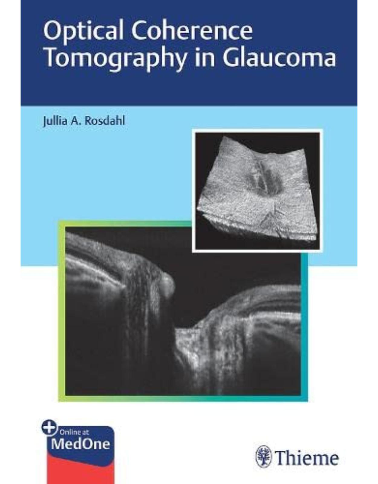
Optical Coherence Tomography in Glaucoma
Livrare gratis la comenzi peste 500 RON. Pentru celelalte comenzi livrarea este 20 RON.
Disponibilitate: La comanda in aproximativ 4-6 saptamani
Autor: Jullia Rosdahl
Editura: Thieme
Limba: Engleza
Nr. pagini: 424
Coperta: Hardcover
Dimensiuni: 17.78 x 1.91 x 25.4 cm
An aparitie: 02/11/2021
Description:
A comprehensive and user-friendly guide on leveraging OCT for the management of glaucoma
Optical coherence tomography (OCT) is a noninvasive diagnostic imaging modality that enables ophthalmologists to visualize different layers of the optic nerve and retinal nerve fiber layer (RNFL) with astounding detail. Today, OCT is an instrumental tool for screening, diagnosing, and tracking the progression of glaucoma in patients. Optical Coherence Tomography in Glaucoma by renowned glaucoma specialist Jullia A. Rosdahl and esteemed contributors is a one-stop, unique resource that summarizes the clinical utility of this imaging technology, from basics to advanced analyses.
The book features 14 chapters, starting with introductory chapters that discuss development of OCT and its applications for visualizing the optic nerve and macula. In chapter 5, case studies illustrate OCT imaging of the optic nerve, RNFL, and macula in all stages of glaucoma, from patients at risk to those with mild, moderate, and severe diseases. The next chapters cover the intrinsic relationship between optic nerve structure and function, the use of structure–function maps, and examples of their relationship, followed by a comparison of commonly used devices and a chapter on artifacts. Anterior segment OCT is covered next, followed by chapters covering special considerations in pediatric glaucomas and in patients with high refractive errors. The final chapters cover innovations in OCT on the horizon including OCT angiography, swept-source OCT, and artificial intelligence.
Key Highlights:
Illustrative case examples provide firsthand clinical insights on how OCT can be leveraged to inform glaucoma treatment.
In-depth guidance on recognizing and managing artifacts including case examples and key technical steps to help prevent their occurrence.
Pearls on the use of OCT for less common patient scenarios such as pediatric glaucomas and high refractive errors.
Future OCT directions including angiography, swept-source, and the use of artificial intelligence.
This practical resource is essential reading for ophthalmology trainees and ophthalmologists new to using OCT for glaucoma. The pearls, examples, and novel topics in this book will also help experienced clinicians deepen their knowledge and increase confidence using OCT in daily practice.
Table of Contents:
1. Introduction: Practical Guide, OCT for Glaucoma
1.1 Introduction
1.2 Overview of the Guide
1.2.1 Development of OCT
1.2.2 OCT of the Optic Nerve
1.2.3 OCT of the Macula
1.2.4 Illustrative Case Examples
1.2.5 Structure–Function Relationship
1.2.6 Comparison of Common Devices
1.2.7 Artifacts and Masqueraders
1.2.8 Anterior Segment OCT in Glaucoma
1.2.9 Special Considerations: OCT in Childhood Glaucoma
1.2.10 Special Considerations: High
1.2.11 Future Directions: OCT Angiography for Glaucoma
1.2.12 Future Directions: Swept-Source OCT for Glaucoma
1.2.13 Future Directions: Artificial Intelligence Applications
1.3 How to Use the Guide
1.4 Conclusion
2. Development of Optical Coherence Tomography
2.1 Introduction
2.2 Setting the Stage: Lasers Meet Medicine
2.2.1 Light in Flight
2.2.2 Interferometry
2.2.3 The Pivotal Role of Collaboration
2.3 Optical Coherence Tomography: The Debut
2.3.1 OCT versus Ultrasound
2.3.2 How OCTWorks
2.3.3 The First OCT Images
2.3.4 Providing Clinical Value
2.4 Commercialization
2.4.1 From the Bench to the Bedside: The Need for Speed
2.4.2 The First Commercial OCT
2.4.3 OCT Instrument Design
2.5 The Fourier Switch
2.5.1 Spectral Domain OCT
2.5.2 Swept-Source OCT
2.6 OCT-Angiography
2.7 Impact
2.7.1 Financial
2.7.2 Scientific and Clinical
3. Optical Coherence Tomography of the Optic Nerve
3.1 Introduction
3.1.1 Glaucoma Imaging
3.1.2 Optical Coherence Tomography
3.2 OCT Output
3.2.1 Overview
3.2.2 OCT Interpretation
3.3 Utility of OCT in Glaucoma Management
3.3.1 Reproducibility
3.3.2 Reliability
3.3.3 Progression Analysis
3.3.4 Diagnostic Accuracy
3.3.5 Correlation with Patient-Centered Outcomes
3.4 Limitations and Pitfalls
3.4.1 Common Artifacts
3.4.2 Pitfalls
3.4.3 Green and Red Disease
3.4.4 Limitations
3.5 Future Directions
3.6 Conclusions
4. Optical Coherence Tomography of the Macula
4.1 Retinal Imaging for Glaucoma
4.1.1 Glaucoma and Retinal Ganglion Cells
4.1.2 RGCs and the Macula
4.2 OCT Image of the Macula
4.2.1 Devices and Segmentation
4.2.2 Correlation between Visual Field and Macular OCT
4.2.3 Complementing Peripapillary RNFL Scans with Macular OCT Scans
4.2.4 Asymmetry Analysis
4.3 Macular OCT and Glaucoma Management
4.3.1 Diagnosis and Progression
4.3.2 Barriers to Proper OCT Interpretation
4.3.3 Practical Uses of Macular OCT
4.4 Artifacts
4.4.1 Vitreous Traction/Adherence
4.4.2 Cystic Changes
4.4.3 Epiretinal Membranes
4.4.4 Retinal Atrophy
4.4.5 Other Macular Pathologies
4.4.6 Myopia
4.4.7 Active Uveitis
4.4.8 Acquisition Artifacts
4.5 OCT Angiography
4.6 Conclusions
5. Illustrative Case Examples
5.1 Overview
5.2 Early (Preperimetric) Glaucoma
5.3 Mild-to-Moderate Glaucoma
5.4 Severe Glaucoma
5.4.1 Severe Stage Glaucoma, due to Significant Visual Field Constriction
5.5 Glaucoma that Is Progressing
5.6 Conclusions
6. Structure–Function Relationship
6.1 Introduction
6.2 Mapping Structural to Functional Loss in Glaucoma
6.2.1 Does OCT Damage Precede Visual Field Loss in Glaucoma?
6.2.2 How Are Structural Changes Linked to Functional Changes in Glaucoma?
7. Comparison of Common Devices
7.1 Retinal Nerve Fiber Layer Thickness
7.1.1 Cirrus 6000 (Carl Zeiss Meditec AG, Jena, Germany)
7.1.2 Spectralis (Heidelberg Engineering GmbH, Heidelberg, Germany)
7.1.3 Avanti RTVue XR (Optovue, Inc., Fremont, CA, USA)
7.1.4 3D OCT (Topcon Corporation, Tokyo, Japan)
7.2 Optic Nerve Head
7.2.1 Cirrus 6000 (Carl Zeiss Meditec AG, Jena, Germany)
7.2.2 Spectralis (Heidelberg Engineering, Inc., Heidelberg, Germany
7.2.3 Avanti RTVue XR (Optovue, Inc., Fremont, CA, USA)
7.2.4 3D OCT (Topcon Corporation, Tokyo, Japan)
7.3 Macula
7.3.1 Cirrus 6000 (Carl Zeiss Meditec AG, Jena, Germany)
7.3.2 Spectralis (Heidelberg Engineering, Inc., Heidelberg, Germany
7.3.3 Avanti RTVue XR (Optovue, Inc., Fremont, CA, USA)
7.3.4 3D OCT (Topcon Corporation, Tokyo, Japan)
7.4 Progression Analysi
7.4.1 Cirrus 6000 (Carl Zeiss Meditec AG, Jena, Germany)
7.4.2 Spectralis (Heidelberg Engineering, Inc., Heidelberg, Germany
7.4.3 Avanti RTVue XR (Optovue, Inc., Fremont, CA, USA)
7.4.4 3D-OCT (Topcon Corporation, Tokyo, Japan)
8. Artifacts and Masqueraders
8.1 Incidence of Artifacts in OCT Imaging
8.2 Etiologies of Peripapillary RNFL OCT Artifacts
8.2.1 Artifacts from Errors in Scan Acquisition
8.2.2 Artifacts in Boundary Segmentation
8.2.3 Artifacts due to Ocular Pathology Unrelated to Glaucoma
8.2.4 Artifacts due to Differences in OCT Machines
8.3 Etiologies of ONH and Macula Artifacts
8.3.1 Bruch’s Membrane Opening– Minimum Rim Width Artifacts
8.3.2 Macular Asymmetry Analysis Artifacts
8.4 OCT Diseases
8.4.1 Red Disease
8.4.2 Green Disease
8.5 A Relevant Summary of OCT Artifacts for Technicians
8.6 Future Directions
8.6.1 Three-Dimensional Parameters
8.6.2 OCT Angiography
9. Anterior Segment Optical Coherence Tomography in Glaucoma
9.1 Introduction to Anterior- Segment OCT
9.2 Different AS-OCT Modalities and Systems
9.3 AS-OCT Identification of Anterior Segment Structures and Parameters
9.4 AS-OCT in Different Glaucomatous Conditions
9.4.1 Primary Angle Closure Suspect (PACS)
9.4.2 Primary Angle Closure Glaucoma (PACG)
9.4.3 AS-OCT Following Laser Peripheral Iridotomy (LPI)
9.4.4 AS-OCT Following Dilation
9.4.5 AS-OCT Following Lens Extraction
9.4.6 Other Glaucomatous Conditions
9.5 AS-OCT in Postoperative Care
9.6 AS-OCT in Additional Glaucoma-Related Situations
9.7 Conclusions
10. Special Considerations: OCT in Childhood Glaucoma
10.1 Introduction
10.2 Handheld and Portable OCT Imaging Modalities
10.3 Image Acquisition
10.3.1 How OCT Can be Used during Examination Under Anesthesia
10.3.2 Tabletop OCT for Children
10.3.3 Structural Considerations for Image Acquisition
10.4 Interpreting OCT Images in Pediatric Glaucoma
10.4.1 Optical and Anatomic Considerations for Image Acquisition and Interpretation
10.4.2 Comparing to a Normative Database
10.4.3 OCT Changes in Childhood Glaucoma
10.4.4 Postoperative Changes in OCT
10.5 Pitfalls and Masqueraders of Pediatric Glaucoma
10.6 Imaging Guidelines and Recommended Frequency of Imaging
11. Special Considerations: High Refractive Errors
11.1 Introduction
11.2 Technical Issues of OCT that Need to be Considered in High Myopia
11.2.1 Scan-Circle Size: Ocular Magnification Error
11.2.2 Scan-Circle Size: Pathologies Influencing the Scan Circle
11.2.3 Scan-Circle Location: Effect of Major Vessels in Tilted Discs
11.2.4 Normative Database
11.2.5 Segmentation Errors
11.3 Glaucoma Diagnosis in High Myopia
11.3.1 Macular Parameters for Glaucoma Diagnosis
11.3.2 Neuroretinal Rim Parameters for Glaucoma Diagnosis
11.3.3 All at One Glance: 3D Wide-Field Map in SS-OCT
12. Future Directions: Optical Coherence Tomography Angiography for Glaucoma
12.1 Introduction
12.1.1 The Role of Ocular Blood Flow in Glaucoma
12.1.2 Optical Coherence Tomography Angiography Technology
12.2 OCTA of the Optic Nerve Head and Peripapillary Microvasculature
12.2.1 OCTA of the Optic Nerve Head Microvasculature in Normal Eyes
12.2.2 OCTA of the Optic Nerve and Peripapillary Microvasculature in Glaucoma
12.3 OCTA of the Macular Microvasculature
12.3.1 OCTA of the Normal Macular Microvasculature
12.3.2 OCTA of Macular Microvasculature in Glaucoma
12.4 OCTA of the Choroid
12.4.1 OCTA of the Normal Choroid
12.4.2 OCTA of the Choroid in Glaucoma
12.5 OCTA in the Future
13. Future Directions: Swept-Source OCT for Glaucoma
13.1 Introduction
13.2 Imaging of Intraocular Structures Using SS-OCT in Glaucoma
13.2.1 Anterior Segment Application of SS-OCT in Glaucoma
13.2.2 Analysis of Macular and Peripapillary Retina by SS-OCT
13.2.3 Choroidal Application of SS-OCT in Glaucoma
13.2.4 Optic Nerve Head Application of SS-OCT in Glaucoma
13.2.5 Lamina Cribrosa Application of SS-OCT in Glaucoma
13.3 Future Research in SS-OCT for Evaluation of Glaucoma
13.4 Conclusions
14. Future Directions: Artificial Intelligence Applications
14.1 Introduction
14.2 Artificial Intelligence
14.2.1 Development of a Deep Learning Algorithm
14.2.2 Deep Learning Algorithms to Diagnose Glaucoma on SD-OCT
14.2.3 Deep Learning Algorithms Trained to Assess Color Fundus Photos Using SD-OCT
14.2.4 Limitations
14.2.5 Future Directions
Index
Read Less
| An aparitie | 02/11/2021 |
| Autor | Jullia Rosdahl |
| Dimensiuni | 17.78 x 1.91 x 25.4 cm |
| Editura | Thieme |
| Format | Hardcover |
| ISBN | 9781684202478 |
| Limba | Engleza |
| Nr pag | 424 |






Clientii ebookshop.ro nu au adaugat inca opinii pentru acest produs. Fii primul care adauga o parere, folosind formularul de mai jos.