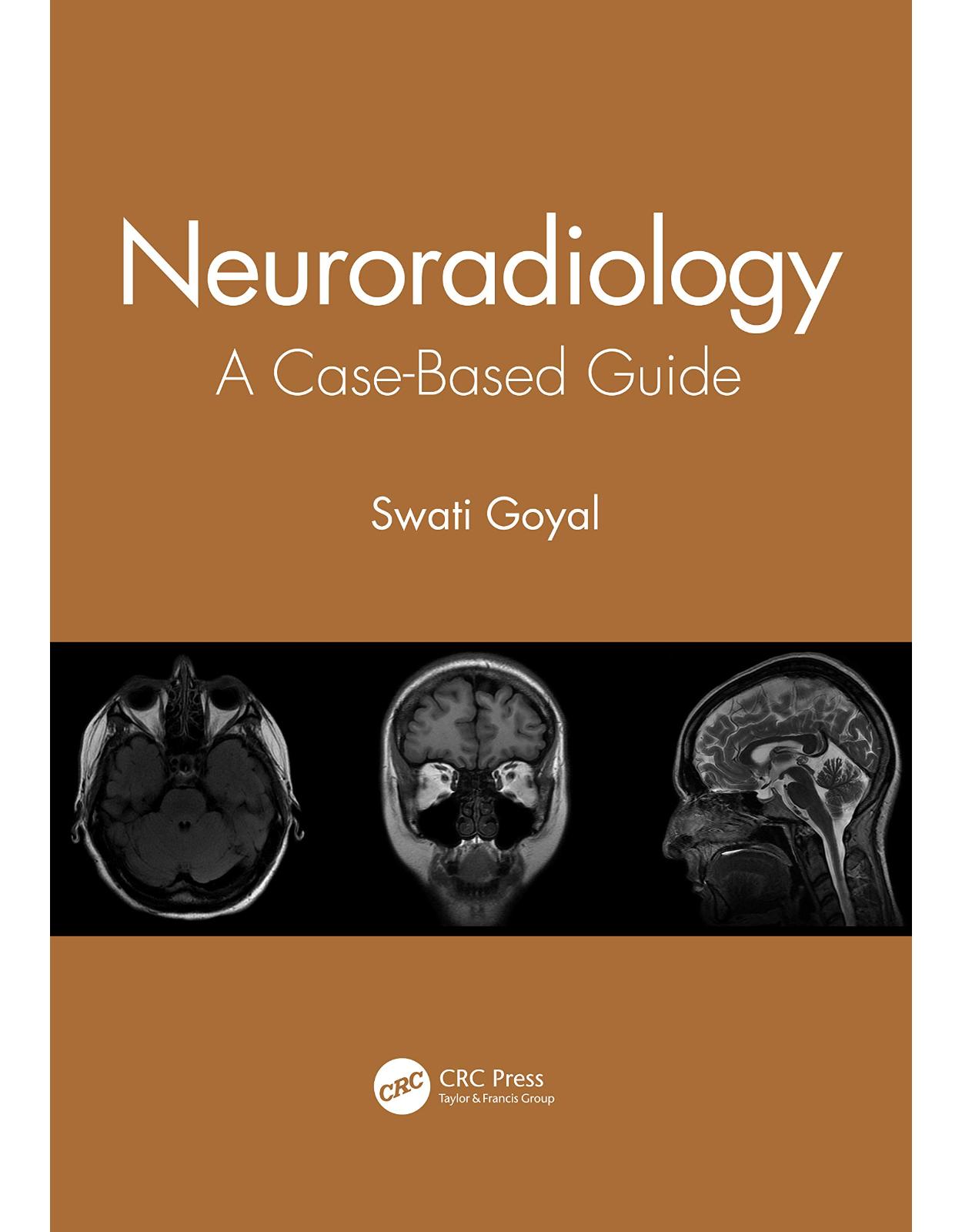
Neuroradiology: A Case-Based Guide
Livrare gratis la comenzi peste 500 RON. Pentru celelalte comenzi livrarea este 20 RON.
Disponibilitate: La comanda in aproximativ 4-6 saptamani
Autor: Swati Goyal
Editura: CRC Press
Limba: Engleza
Nr. pagini: 222
Coperta: Paperback
Dimensiuni:
An aparitie: 23 Oct. 2020
Description:
This book covers the complete gamut of neuroradiology cases, including normal anatomy, pitfalls, and artifacts across the brain and spine in a single volume, enriched with high-resolution images that support the interpretation of CT and MRI images of the brain, spine, head, and neck. It includes case studies commonly encountered in clinical practice, in addition to normal anatomy, that prepare the reader for the challenges in the clinical setting. Each case study discusses the clinical history, relevant imaging findings, differential diagnosis, and management, serving as a helpful read for trainee radiologists, neurophysicians, neurosurgeons, and CT/MRI technicians, along with physicians interested in medical imaging. Key Features Provides a succinct overview of normal variants with case studies structured into thematic chapters Serves as a basic accompaniment for radiology residents, fellows, practicing radiologists, neurophysicians, neurosurgeons, emergency medicine practitioners, trainee and practicing radiographers, and those studying for Board exams Highlights the relevance of artificial intelligence in clinical practice
Table of Contents:
1. PART I THE BRAIN
2. 1 Normal Brain Development and Congenital Malformations
3. Case Studies
4. Chiari Malformations
5. Lissencephaly
6. Gray Matter Heterotopias
7. Schizencephaly (Split Brain)
8. Holoprosencephaly
9. Corpus Callosum Agenesis (CCA)
10. Hemimegalencephaly
11. Dyke-Davidoff-Masson Syndrome
12. Perisylvian Syndrome (Opercular Syndrome, Perisylvian Polymicrogyria, Worster-Drought Syndrome)
13. Hypoxic-Ischemic Encephalopathy (HIE/Hypoxic-Ischemic Injury)
14. Porencephalic Cyst
15. Dandy-Walker Malformation
16. Joubert’s Syndrome
17. Suggested Reading
18. 2 Vascular Anatomy
19. Internal Carotid Artery – Segments and Their Branches
20. Variants of ICA
21. Aberrant ICA
22. Normal Variants
23. Cerebral Arteries
24. Anterior Cerebral Artery (ACA)
25. Middle Cerebral Artery (MCA)
26. Posterior Cerebral Artery (PCA)
27. Vertebrobasilar System
28. Vertebral Artery
29. Basilar Artery
30. Venous Anatomy
31. Dural Sinuses
32. Superficial Cortical Veins
33. Deep Cerebral Veins
34. Stroke
35. Stroke in Children
36. Stroke Mimics
37. Intracranial Aneurysms
38. Etiology
39. Intracranial Vascular Malformations
40. Case Studies
41. ICA Aneurysm
42. Ruptured ACom Aneurysm
43. Basilar Tip Aneurysm
44. Cavernoma
45. Developmental Venous Anomaly (DVA)/Venous Angioma
46. Arteriovenous Malformations (AVMs)
47. Cirsoid Aneurysm of Scalp (Plexiform Angioma)
48. Anterior Cerebral Artery Infarct
49. Middle Cerebral Artery (MCA) Infarct
50. Posterior Cerebral Artery Infarct
51. Artery of Percheron Infarct
52. Posterior Inferior Cerebellar Artery (PICA) Infarct – Lateral Medullary Syndrome (Wallenberg Syndrome)
53. Watershed (Border Zone) Infarct
54. Lacunar Infarct
55. Cerebral Venous Thrombosis
56. Venous Infarct with Dural Sinus Thrombosis
57. Moyamoya Disease
58. Cerebral Autosomal Dominant Arteriopathy with Subcortical Infarcts and Leukoencephalopathy (CADASIL)
59. Posterior Reversible Encephalopathy Syndrome (PRES)
60. Cortical Laminar Necrosis
61. Imaging
62. Fetal Origin of Posterior Cerebral Artery (PCA)
63. Persistent Trigeminal Artery (PTA)
64. Suggested Reading
65. 3 Intracranial Hemorrhage (ICH)
66. Perinatal Hemorrhage
67. Preterm Infants
68. Germinal Matrix Hemorrhage (GMH)
69. Term Infants
70. Traumatic Extracranial Hemorrhage
71. Traumatic Intracranial Hemorrhage
72. Non-Traumatic Intracranial Hemorrhage
73. Adults
74. Intraventricular Hemorrhage (IVH)
75. Subdural Hemorrhage (SDH) vs. Extradural Hemorrhage (EDH) and Cerebral Herniation
76. Case Studies
77. Subdural Hemorrhage (SDH)
78. Epidural Hemorrhage
79. Subarachnoid Hemorrhage (SAH)
80. Hemorrhagic Parenchymal Contusions
81. Hypertensive Hemorrhage
82. Diffuse Axonal Injury (DAI)
83. Management
84. Non-Accidental Injury (NAI) (Battered Child Syndrome, Shaken Infant Syndrome)
85. Depressed Skull Fracture
86. Leptomeningeal Cyst (Growing Skull Fracture)
87. Suggested Reading
88. 4 Radiology of Brain Tumors
89. Analytical Approach
90. Age Distribution
91. Location
92. Edema
93. Midline Crossing Lesions
94. Multifocal/Multiple Tumors
95. Signal Characteristics
96. Tumors with High Density on CT Scan
97. Fat
98. Calcification
99. Low SI on T2WI
100. High Signal Intensity on T1
101. Enhancement Pattern
102. Little/No Enhancement
103. Ring Enhancement
104. Recent Additions to the WHO Classification of Tumors
105. MVNT (Multinodular Vacuolating Neuronal Tumor) of the Cerebrum
106. Diffuse Leptomeningeal Glioneuronal Tumor
107. Tumor Mimics
108. Imaging
109. Long-Term Complications of Chemo-/Radiotherapy
110. Classification
111. Supratentorial Tumors
112. Secondary Brain Tumors (Metastasis)
113. Infratentorial Tumors
114. Case Studies
115. Glioblastoma Multiforme
116. Oligodendroglioma
117. Meningioma
118. Cerebello-Pontine Angle (CPA) Schwannoma (Acoustic Neuroma/ Vestibular Schwannoma)
119. Pituitary Adenoma
120. Pituitary Apoplexy
121. Craniopharyngioma
122. Epidermoid Cyst
123. Central Neurocytoma
124. Medulloblastoma
125. Pineoblastoma
126. Choroid Plexus Papilloma
127. Colloid Cyst
128. Rathke’s Cleft Cyst
129. Hemangioblastoma
130. Primary CNS Lymphoma (PCNSL)
131. Brainstem Glioma
132. Dysembryoplastic Neuroepithelial Tumors (DNET)
133. Primitive Neuroectodermal Tumor (PNET)
134. Intracranial Lipoma
135. Ruptured Intracranial Dermoid Cyst
136. CNS Metastasis
137. Suggested Reading
138. 5 Infections and Inflammatory Diseases
139. Congenital Brain Infections
140. Acute Pyogenic Meningitis
141. Subdural Empyema with Cerebral Abscess
142. Otogenic Brain Abscess
143. Cerebral Abscess
144. Neurocysticercosis (NCC)
145. Tuberculoma
146. Herpes Encephalitis
147. Idiopathic Hypertrophic Pachymeningitis
148. Acute Necrotizing Encephalitis
149. Suggested Reading
150. 6 Disorders Affecting White and Gray Matter: Normal Myelination
151. Classification
152. Demyelinating Disorders
153. Dysmyelination Disorders (Leukodystrophies)
154. Metachromatic Leukodystrophy (MLD)
155. Krabbe Disease (Globoid Leukodystrophy)
156. Alexander Disease
157. Canavan’s Disease
158. Pelizaeus-Merzbacher Disease
159. Phenylketonuria (PKU)
160. Homocystinuria
161. Glutaric Aciduria
162. Methylmalonic Acidemia (MMA)
163. Disorders Primarily Affecting Gray Matter (GM)
164. Tay-Sachs Disease
165. Mucopolysaccharidoses (MPS)
166. MPS 1 (Hurler’s Syndrome)
167. Glycogen Storage Diseases
168. Mucolipidoses and Fucosidoses
169. Disorders Affecting Both Gray and White Matter
170. Leigh’s Syndrome
171. Myopathy, Encephalopathy, Lactic Acidosis, and Stroke-Like Episodes (MELAS) Syndrome
172. Basal Ganglia Disorders
173. Primary NeuroDegenerative Conditions
174. Etiology
175. Case Studies
176. Multiple Sclerosis
177. Adrenoleukodystrophy (ALD)
178. Alzheimer’s Disease
179. Fahr’s Disease (Familial Brain Calcification)
180. Hallervordern-Spatz Disease (Pantothenate Kinase-Associated Neurodegeneration, PKAN)
181. Non-Ketotic Hyperglycemia-Hemichorea-Hemiballismus (NKHHH Diabetic Striatrophy)
182. Suggested Reading
183. 7 CSF Circulation and Disorders
184. Sources of CSF Production
185. CSF Drainage
186. CSF Absorption
187. Ventricles
188. Lateral Ventricles
189. CSF Flow Pathway
190. Cisterns
191. CSF Flow Artifacts
192. Normal Variants
193. Asymmetric Lateral Ventricles
194. Case Studies
195. Hydrocephalus
196. Normal Pressure Hydrocephalus (NPH)
197. Benign Enlargement of Subarachnoid Spaces in Infancy (BESSI)
198. Suggested Reading
199. 8 Phakomatoses (Neurocutaneous Syndromes)
200. Other Neurocutaneous Syndromes
201. Case Studies
202. Neurofibromatosis Type 1 (NF1, von Recklinghausen Disease)
203. Neurofibromatosis Type 2
204. Segmental Neurofibromatosis
205. Tuberosus Sclerosis (TS, Bourneville Disease)
206. Sturge-Weber Syndrome (Encephalotrigeminal Angiomatosis)
207. Von Hippel-Landau (VHL) Syndrome
208. Suggested Reading
209. 9 Abnormal Skull
210. Congenital Lesions
211. Lacunar Skull (Luckenschadel)
212. Sinus Pericranii
213. Intracranial Calcifications
214. Skull Erosions
215. Hyperostosis
216. Skull Base and Its Foramina
217. Cranial Nerves
218. Case Studies
219. Craniosynostosis (Craniostenosis, Sutural Synostosis, or Cranial Dysostosis)
220. Persistent Metopic Suture (Metopism/Sutura Frontalis Persistans)
221. Frontoethmoidal (Sincipital) Encephalocele
222. Bell’s Palsy (Idiopathic Peripheral Facial Paralysis)
223. Management
224. Skull Base (Clival) Chordoma
225. Suggested Reading
226. PART II: THE SPINE
227. 10 Craniovertebral Junction Anomalies
228. Craniometry
229. External Craniocervical Ligaments
230. Internal Craniocervical Ligaments
231. Anomalies of Occiput
232. Anomalies of Atlas
233. Anomalies of Axis
234. Congenital Malformations
235. Acquired Disorders
236. Trauma
237. Basilar Invagination
238. Os Odontoideum
239. Imaging and DDs
240. Ancillary
241. Odontoid Fracture
242. Suggested Reading
243. 11 Congenital Anomalies
244. Normal Anatomy
245. Lipomyelocele
246. Diastematomyelia
247. Caudal Regression Syndrome
248. Suggested Reading
249. 12 Acquired Diseases
250. Normal Anatomy
251. Ligaments
252. Cervical Spine
253. Thoracolumbar Spine
254. MRI Features
255. Spinal Lesions
256. Disc Herniation with Extrusion
257. Vertebral Hemangioma
258. Pott’s Spine (Tuberculous Spondylitis)
259. Transverse Myelitis
260. Burst Fractures
261. Spondylolisthesis
262. Brachial Plexus Injury with Traumatic Pseudomeningocele
263. Spinal Schwannoma (Neurinoma/Neurilemmoma)
264. Dural Ectasia
265. Spinal Dural AVF (Arteriovenous Fistula)
266. Ankylosing Spondylitis (AS)
267. Hangman’s Fracture (Traumatic Spondylolisthesis of Axis)
268. Vertebral Metastasis with Cord Compression
269. Tarlov Cyst (Perineural Cyst)
270. Myxopapillary Ependymoma
271. Primary Lymphoma of Spine
272. Suggested Reading
273. PART III: THE HEAD AND NECK
274. 13 Orbit
275. Boundaries of the Orbit
276. Major Apertures
277. Globe (Ocular Space)
278. Orbit and Optic Nerve Sheath Complex
279. Conal-Intraconal Area
280. Extraconal Area
281. Orbital Appendages
282. Case Studies
283. Graves’ Ophthalmopathy
284. Orbital Pseudotumor
285. Optic Neuritis
286. Tolosa-Hunt Syndrome
287. Retinoblastoma
288. Optic Nerve Glioma
289. Choroidal Melanoma (Uveal Melanoma)
290. Orbital Blow-Out Fracture
291. Cavernous Hemangioma
292. Carotid-Cavernous Fistula
293. Suggested Reading
294. 14 Neck
295. Neck Spaces of Supra- and Infrahyoid Region and Fascial Layers
296. Visceral Space
297. Retropharyngeal Space (RPS)
298. Prevertebral Space (PVS)
299. Perivertebral Space
300. Parapharyngeal Space (PPS)
301. Pharyngeal Mucosal Space (PMS)
302. Carotid Space (CS)/Post-Styloid PPS, Carotid Sheath
303. Masticator Space (MS)
304. Parotid Space
305. Buccal Space
306. Sublingual Space (SLS)/Floor of Mouth
307. Submandibular Space (SMS)
308. Vascular Structures of the Neck
309. Branches of the External Carotid Artery
310. Salivary Glands
311. Lymph Nodes of the Neck
312. Larynx
313. Thyroid
314. Case Studies
315. Thyroglossal Duct Cyst (TGDC)
316. Branchial Cleft Cyst (BCC)
317. Other Types of Branchial Cleft Cysts
318. Cystic Hygroma (Nuchal Lymphangioma)
319. Laryngocele
320. Calcific Longus Colli Tendonitis (Retropharyngeal/Acute Calcific Prevertebral Tendonitis)
321. Retropharyngeal Abscess (RPA)
322. Carotid Body Tumor
323. Laryngeal Carcinoma
324. Supraglottic Carcinoma
325. Glottic Carcinoma
326. Submandibular Duct Calculi (Sialolithiasis)
327. Pleomorphic Adenoma
328. Ranula
329. Suggested Reading
330. 15 Ear, Nose, and Paranasal Sinus
331. Ear
332. Congenital Anomalies
333. Pulsatile Tinnitus
334. Nasal Cavity and Paranasal Sinuses
335. Maxillary Sinus
336. Ethmoid Sinus
337. Frontal Sinus
338. Sphenoid Sinus
339. Normal Anatomical Variations
340. Chronic Otitis Media with Mastoiditis
341. Cholesteatoma
342. Cholesterol Granuloma
343. Antrochoanal Polyp
344. Frontoethmoidal Mucocele
345. Osteomeatal Unit (OMU) Pattern Obstructive Sinusitis
346. Invasive Fungal Sinusitis
347. Frontal (Ivory) Osteoma
348. Juvenile Ossifying Fibroma
349. Esthesioneuroblastoma (ENB, Olfactory Neuroblastoma)
350. Nasopharyngeal Carcinoma
351. Juvenile Nasopharyngeal Angiofibroma
352. Ameloblastoma (Adamantinoma of the Jaw)
353. Dentigerous Cyst (Follicular Cyst)
354. Thornwaldt Cyst
355. Rhinolith (Nasal Calculi)
356. Suggested Reading
357. 16 Miscellaneous
358. Epilepsy
359. Types
360. Focal (Partial)
361. Generalized
362. Mesial Temporal Sclerosis (MTS) (Hippocampal Sclerosis)
363. Empty Sella
364. AI in Radiology
365. What AI Can Do
366. What AI Cannot Do
367. Suggested Reading
368. Index
| An aparitie | 23 Oct. 2020 |
| Autor | Swati Goyal |
| Editura | CRC Press |
| Format | Paperback |
| ISBN | 9780367903190 |
| Limba | Engleza |
| Nr pag | 222 |

