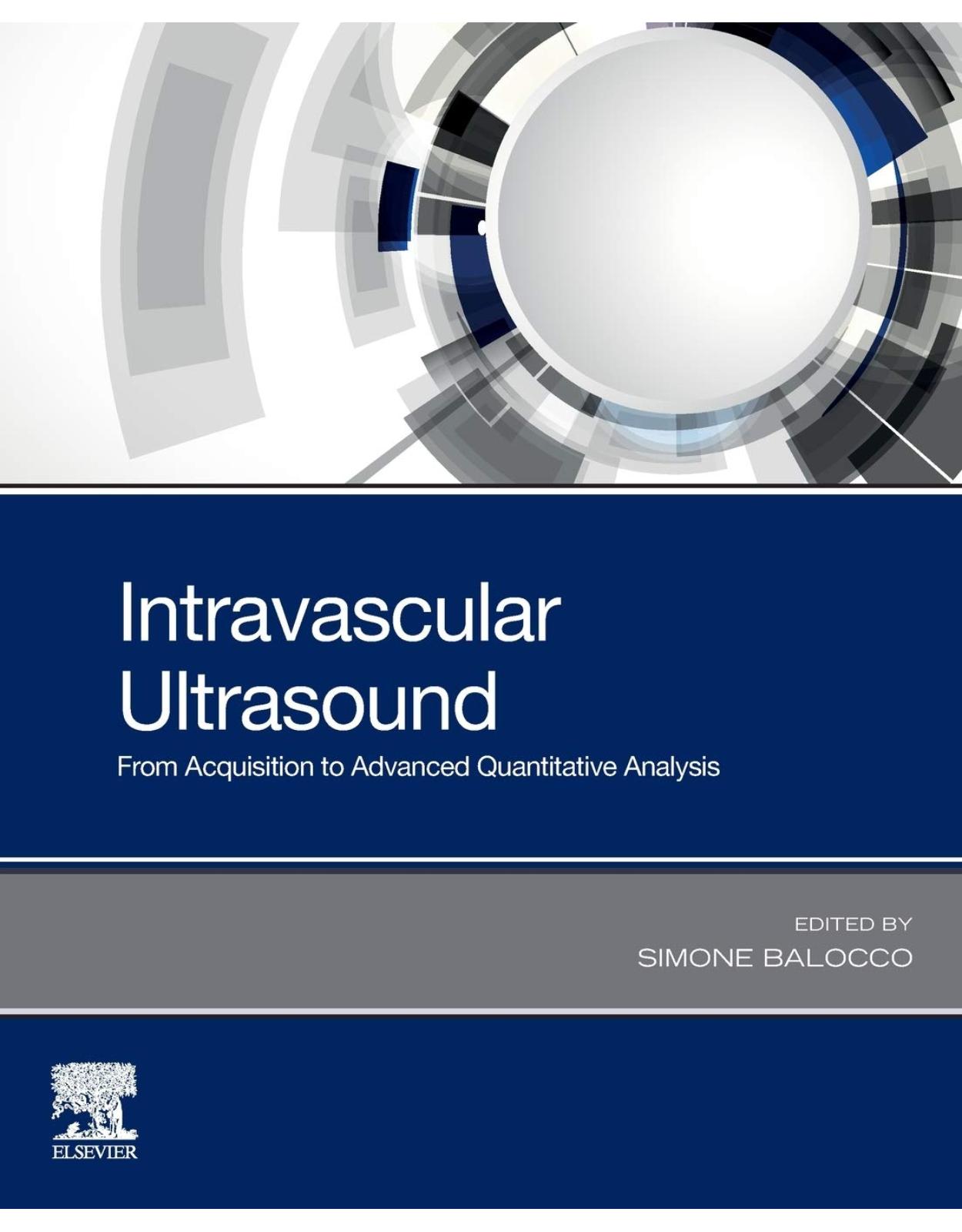
Intravascular Ultrasound: From Acquisition to Advanced Quantitative Analysis
Livrare gratis la comenzi peste 500 RON. Pentru celelalte comenzi livrarea este 20 RON.
Disponibilitate: La comanda in aproximativ 4-6 saptamani
Autor: Simone Balocco
Editura: Elsevier
Limba: Engleza
Nr. pagini: 218
Coperta: Paperback
Dimensiuni: 19.05 x 1.17 x 23.5 cm
An aparitie: 23 Jun. 2020
Description:
Intravascular Ultrasound: From Acquisition to Advanced Quantitative Analysis covers topics of the whole imaging pipeline, ranging from the definition of the clinical problem and image acquisition systems to image processing and analysis, including the assisted clinical-decision making procedures and treatment planning (stent deployment and follow up). Atherosclerosis, a disease of the vessel wall that produces vessel narrowing and obstruction, is the major cause of cardiovascular diseases, such as heart attack or stroke. This book covers all aspects of this imaging tool that allows for the visualization of internal vessel structures and the quantification and characterization of coronary plaque.
Provides an introduction to the clinical workflow and current challenges in endovascular interventions
Presents a review of the state-of-the-art methodologies in IVUS imaging and their applications
Includes a rich analysis of the current and potential future connections between the academic, clinical and industrial fields
Table of Contents:
Chapter 1: Introduction
Section I: Clinical Ivus
Chapter 2: Clinical Utility of Intravascular Ultrasound
1. Introduction
2. Basic Principles of Imaging Acquisition
3. IVUS in Clinical Practice
3.1. IVUS in Percutaneous Transluminal Coronary Angioplasty
3.2. IVUS in the Bare Metal Stent Era
3.3. IVUS in the Drug-Eluting Stent Era
4. IVUS Applications
4.1. IB-IVUS
4.2. VH-IVUS
4.3. iMap-IVUS
4.4. NIRS-IVUS
4.5. Contrast-Enhanced IVUS
5. IVUS Assessment of Plaque Progression
6. IVUS in Complex Coronary Lesions
6.1. Left Main Coronary Artery Lesions
6.2. Other Coronary Lesions
6.3. Calcified Lesions
6.4. Coronary Chronic Total Occlusion
7. IVUS Pitfalls
8. IVUS Complications
9. Future Perspectives
References
Chapter 3: Convenience of Intravascular Ultrasound in Coronary Chronic Total Occlusion Recanalizatio
1. Innovation Chronology on CTO Coronary Intervention Guided by IVUS
2. Why and When IVUS Is Used in CTO Recanalization
2.1. Anterograde Approach to CTO Recanalization
2.1.1. IVUS to define the proximal edge and validate the true lumen access
2.1.2. IVUS to delimit true lumen situation versus guidewire position along with the CTO stenosis un
2.1.3. IVUS to size and optimize the stent
2.2. IVUS in Retrograde CTO Recanalization
2.2.1. IVUS in reverse recanalization
2.2.2. IVUS in reverse CART
2.2.3. IVUS in bailout
2.2.4. Size and optimize the stent
3. Case Examples
3.1. Case 1 Example
Anterograde RCA CTO
Introduction
3.2. Case 2 Example
Anterograde LAD CTO
Introduction
3.3. Case 3 Example
Ostial LAD CTO procedure guided from diagonal IVUS
Introduction
3.4. Case 4 Example
Calcified CTO in LAD
Introduction
3.5. Case 5 Example
LAD CTO, anterograde wired in false lumen
Introduction
3.6. Case 6 Example
LM severe stenosis and LAD CTO
Introduction
3.7. Case 7 Example
LAD CTO, bailout retrograde from RCA
Introduction
References
Chapter 4: Intracardiac Ultrasound
1. Introduction
2. ICE Devices and Case Studies
3. Rotational System
4. Phased Array System
5. Position and Image Examples of Phased Array System3
6. Case Examples
7. Patent Foramen Ovale7
8. Atrial Septum Defect
9. Hypertrophic Obstructive Cardiomyopathy8
10. Left Atrial Appendage Closure9
11. 3D/4D Developments (Phased Array Only)
12. Transseptal Puncture
13. Cryoablation; Positioning of the Cryoballoon
14. PFO Closure
References
Chapter 5: Quantitative Virtual Histology for In Vivo Evaluation of Human Atherosclerosis-A Plaque B
Summary
List of Abbreviations
1. Introduction
2. Methodology
3. Results
4. Discussion
References
Section II: Ivus Image Analysis
Chapter 6: A State-of-the-Art Intravascular Ultrasound Diagnostic System
1. Introduction
1.1. Overview
1.2. Procedure of IVUS Observation
1.2.1. Preparation
1.2.2. Observation
1.2.3. Catheter removal
2. IVUS Imaging Catheter
2.1. Concept
2.2. Requirements for Catheter
2.2.1. Trackability
2.2.2. Pushability
2.2.3. Crossability
2.2.4. Visibility
2.2.5. Safety
2.3. Requirements for Ultrasound Probe
2.3.1. Radial imaging
2.3.2. Longitudinal imaging
2.3.3. Ultrasound transmission
3. System Configuration
3.1. Motor Drive Unit
3.2. Console
4. Signal Processing
4.1. Image Construction
4.2. High-Frequency IVUS
5. Concluding Remarks
References
Chapter 7: Multimodality Intravascular Imaging Technology
1. Introduction
2. Hybrid IVUS-OCT Imaging Technology
3. Near-Infrared Spectroscopy
4. Intravascular Photoacoustics
5. Near-Infrared Fluorescence
6. Fluorescence Lifetime Imaging
7. Multimodality Imaging: Clinical Outlook
References
Chapter 8: Quantitative Assessment and Prediction of Coronary Plaque Development Using Serial Intrav
1. Introduction
1.1. Segmentation and Registration Approaches
1.2. Quantitative Assessment and Prediction
1.3. Study Background
2. Graph-Based IVUS Segmentation
2.1. Optimal Graph Search: LOGISMOS
2.2. Graph and Cost-Function Design
2.3. Advanced Editing: Just Enough Interaction
3. Baseline/Follow-Up Registration
3.1. 3D Correspondence Graph
3.2. Feature- and Morphology-Based Similarity
3.3. Finding the Start and End Frame Pairs
3.4. Validation Results
4. Morphology Assessment: Remodeling
4.1. Progression and Regression Groups
4.2. Plaque and Adventitia Development
5. Prediction of Plaque Development
5.1. Plaque Phenotype Definitions
5.2. Features and Classifiers
5.3. Feature Selection for Predictions
5.4. Analysis and Prediction Results
6. Conclusions
Glossary
References
Chapter 9: Training Convolutional Nets to Detect Calcified Plaque in IVUS Sequences
1. Introduction
2. Methodology
2.1. Frame-Based Detection
2.1.1. Convolutional architectures
2.1.2. Training deep calcified plaque detectors
2.2. Improving Temporal Consistency
2.3. Final Selection of Relevant Frames
3. Experiments and Discussion
3.1. Materials
3.2. Evaluation Metrics
3.3. Experiments
3.4. Results
3.5. Qualitative Evaluation
4. Conclusions
References
Chapter 10: Computer-Aided Detection of Intracoronary Stent Location and Extension in Intravascular
1. Introduction
2. Stent Shape Estimation
2.1. State of the Art of Stent Shape Estimation
2.2. Method
2.2.1. Gating
2.2.2. Semantic classification
Classes definition
Pixel-wise descriptor
Semantic classification framework
2.2.3. Stent modeling
Stent shape estimation
Strut detection
2.3. Validation
2.3.1. Material
2.3.2. Experiments on stent modeling
2.4. Discussion
2.4.1. Metallic stents
Bioabsorbable stents
2.5. Conclusion
3. Longitudinal Stent Location
3.1. State of the Art of Longitudinal Stent Location
3.2. Method
3.2.1. Gating and strut detection
3.2.2. Stent presence assessment
3.3. Validation
3.3.1. Material
3.3.2. Experiments on stent presence assessment
3.3.3. Malapposition analysis
3.4. Results
3.4.1. Metallic stents
Bioabsorbable stents
3.5. Discussion
3.6. Conclusion
4. General Conclusion
References
Chapter 11: Real-Time Robust Simultaneous Catheter and Environment Modeling for Endovascular Navigat
1. Introduction
2. Vessel Modeling in SCEM
3. Methods
3.1. Optimization Based on the Preoperative Data
3.2. Uncertainty Estimation
3.3. Real-Time Implementation
4. Results
4.1. Simulation and Robustness Assessment
4.2. Phantom Experiments
5. Conclusion
References
Index
Back Cover
| An aparitie | 23 Jun. 2020 |
| Autor | Simone Balocco |
| Dimensiuni | 19.05 x 1.17 x 23.5 cm |
| Editura | Elsevier |
| Format | Paperback |
| ISBN | 9780128188330 |
| Limba | Engleza |
| Nr pag | 218 |



Clientii ebookshop.ro nu au adaugat inca opinii pentru acest produs. Fii primul care adauga o parere, folosind formularul de mai jos.