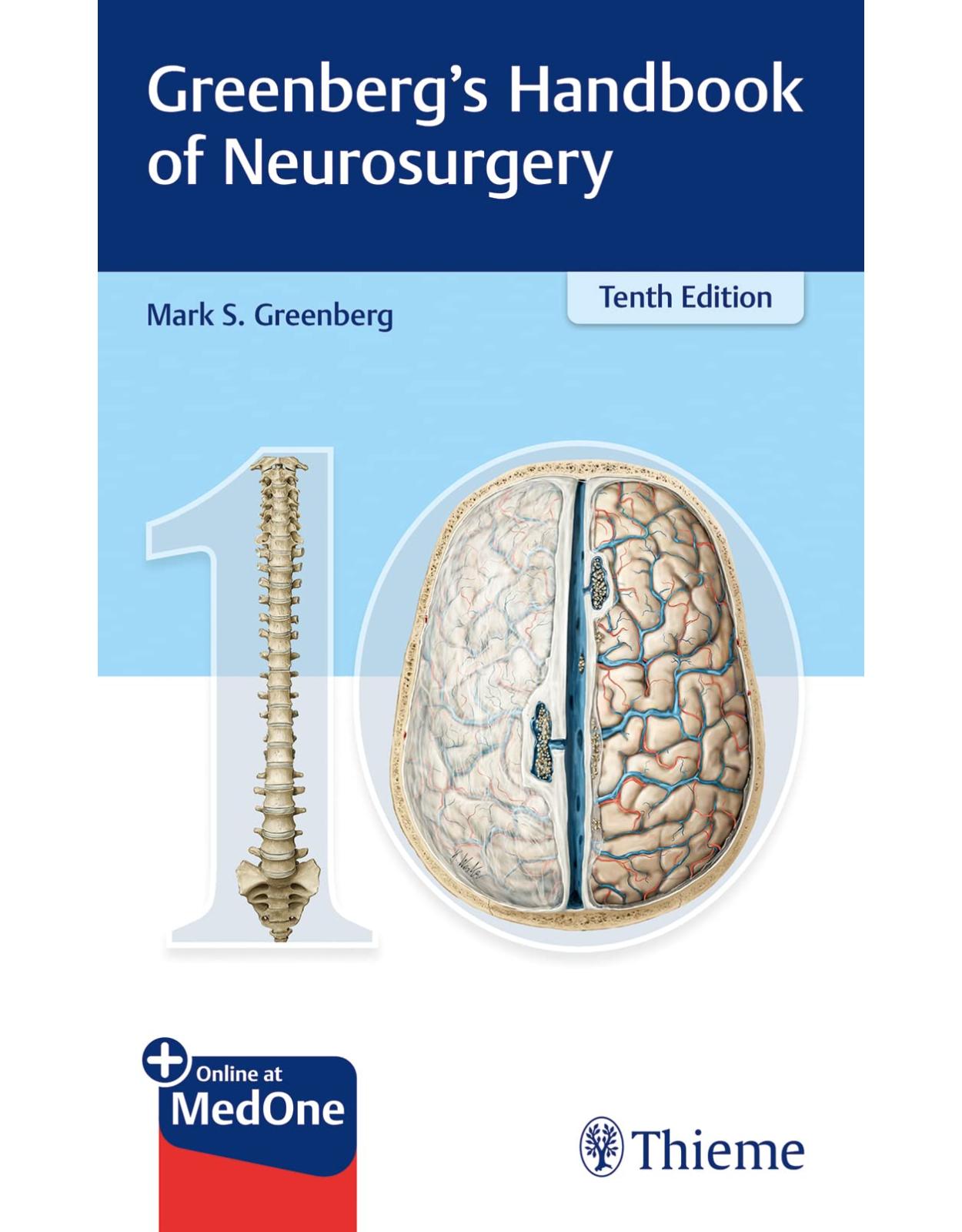
Greenberg’s Handbook of Neurosurgery
Livrare gratis la comenzi peste 500 RON. Pentru celelalte comenzi livrarea este 20 RON.
Disponibilitate: La comanda in aproximativ 4 saptamani
Autor: Mark S. Greenberg
Editura: Thieme
Limba: Engleza
Nr. pagini: 1990
Coperta: Paperback
Dimensiuni: 13.2 x 5.8 x 20.3 cm
An aparitie: 19/04/2023
Description:
The fundamental, one-stop global resource for neurosurgical practice in updated 10th edition
Unlike traditional medical textbooks, the origins of the now legendary Handbook of Neurosurgery by Mark Greenberg took root in the late 1980's in the notes the author kept while taking care of his patients, from his weekly grand rounds presentations, and in the literature he read. Now in its 10th edition, the book that is often referred to as the "bible for neurosurgeons" has grown organically over the years to include more topics of importance to those who provide healthcare to patients with neurologic ailments, and to reflect advances in the understanding and management of the underlying conditions and diseases.
Throughout 23 sections and 116 chapters, the generously illustrated text covers information ranging from pediatrics to geriatrics. The comprehensive, conveniently compact book provides detailed, high-value, and actionable information that can be quickly accessed to enhance patient management, thereby eliminating the need to wade through superfluous material. It is also a perfect study companion for board examination and preparation for the maintenance of certification.
Key Features:
Updated classification and diagnostic criteria of central and peripheral nervous system tumors, based on the most recent WHO classifications
Reworking of numerous sections (including current concepts in pseudotumor cerebri, seizure classification…)
Addition of new chapters (including idiopathic scoliosis, LOVA and tuberculosis of the CNS)
Current principles of nonsurgical and surgical management, including risk factors, indications, diagnostics, prognoses, contraindications, and differential diagnoses
Nearly 500 figures, including new summary flow charts, illustrations, and simplified diagrams for early learners, enhance understanding of material discussed in the text
And, as always, thousands of references!
This unique book encompasses a wide breadth of neurosurgical topics in an easy digestible format, making it an indispensable daily reference for all neurosurgical residents, fellows, neurosurgeons, and allied health practitioners.
Table of Contents:
Section I Anatomy and Physiology
1 Gross Anatomy, Cranial and Spine
1.1 Cortical surface anatomy
1.1.1 Lateral cortical surface
1.1.2 Brodmann’s areas
1.1.3 Medial surface
1.1.4 Somatotopic organization of primary sensory and motor cortex
1.2 Central sulcus on axial imaging
1.3 Surface anatomy of the cranium
1.3.1 Craniometric points
1.3.2 Relation of skull markings to cerebral anatomy
1.3.3 Relationship of ventricles to skull
1.4 Surface landmarks of spine levels
1.5 Cranial foramina and their contents
1.5.1 Summary
1.5.2 Porus acusticus
1.6 Internal capsule
1.6.1 Architectural anatomy
1.6.2 Vascular supply of the internal capsule (IC)
1.7 Cerebellopontine angle anatomy
1.8 Occipitoatlantoaxial-complex anatomy
1.9 Spinal cord anatomy
1.9.1 Dentate ligament
1.9.2 Spinal cord tracts
1.9.3 Dermatomes and sensory nerves
1.10 References
2 Vascular Anatomy
2.1 Cerebral vascular territories
2.2 Cerebral arterial anatomy
2.2.1 General information
2.2.2 Circle of Willis
2.2.3 Anatomical segments of intracranial cerebral arteries
2.2.4 Anterior circulation
2.2.5 Posterior circulation
2.2.6 Fetal carotid-vertebrobasilar anastomoses
2.3 Cerebral venous anatomy
2.3.1 Supratentorial venous system
2.3.2 Posterior fossa venous anatomy
2.4 Spinal cord vasculature
2.5 References
3 Neurophysiology and Regional Brain Syndromes
3.1 Neurophysiology
3.1.1 Blood-brain barrier
3.1.2 Language and speech function
3.1.3 Babinski sign and Hoffmann sign
3.1.4 Bladder neurophysiology
3.2 Regional brain syndromes
3.2.1 Overview
3.2.2 Parietal lobe syndromes
3.2.3 Foster Kennedy syndrome
3.2.4 Cerebellar mutism & syndromes of the posterior fossa
3.2.5 Brainstem and related syndromes
3.2.6 Parinaud’s syndrome
3.3 Jugular foramen syndromes
3.3.1 Applied anatomy
3.3.2 Clinical syndromes
3.4 References
Section II General and Neurology
4 Neuroanesthesia
4.1 ASA classification
4.2 Neuroanesthesia parameters
4.3 Drugs used in neuroanesthesia
4.3.1 Inhalational agents
4.3.2 Intravenous anesthetic agents
4.3.3 Miscellaneous drugs in neuroanesthesia
4.3.4 Paralytics for intubation
4.4 Anesthetic requirements for intraoperative evoked potential monitoring
4.5 Malignant hyperthermia
4.5.1 General information
4.5.2 Presentation
4.5.3 Treatment
4.5.4 Prevention
4.6 References
5 Sodium Homeostasis and Osmolality
5.1 Serum osmolality and sodium concentration
5.2 Hyponatremia
5.2.1 General information
5.2.2 Evaluation of hyponatremia
5.2.3 Symptoms
5.2.4 Syndrome of inappropriate antidiuresis (SIAD)
5.2.5 Syndrome of inappropriate antidiuretic hormone secretion (SIADH)
5.2.6 Cerebral salt wasting
5.3 Hypernatremia
5.3.1 General information
5.3.2 Diabetes insipidus
5.4 References
6 General Neurocritical Care
6.1 Parenteral agents for hypertension
6.2 Hypotension (shock)
6.2.1 Classification
6.2.2 Cardiovascular agents for shock
6.3 Acid inhibitors
6.3.1 Stress ulcers in neurosurgery
6.3.2 Prophylaxis for stress ulcers
6.3.3 Possible increased pneumonia and mortality from altering gastric pH
6.3.4 Histamine2 (H2) antagonists
6.3.5 Gastric acid secretion inhibitors (proton pump inhibitors)
6.3.6 Miscellaneous
6.4 Rhabdomyolysis
6.4.1 Background and pathophysiology
6.4.2 Etiology and epidemiology
6.4.3 Management and treatment
6.5 References
7 Sedatives, Paralytics, Analgesics
7.1 Sedatives and paralytics
7.1.1 Richmond agitation-sedation scale (RASS)
7.1.2 Conscious sedation
7.1.3 Sedation
7.2 Paralytics (neuromuscular blocking agents)
7.2.1 General information
7.2.2 Ultra-short acting paralytics
7.2.3 Short acting paralytics
7.2.4 Intermediate acting paralytics
7.2.5 Reversal of competitive muscle blockade
7.3 Analgesics
7.3.1 General information
7.3.2 Guiding principles
7.3.3 Analgesics for some specific types of pain
7.3.4 Nonopioid analgesics
7.3.5 Opioid analgesics
7.3.6 Adjuvant pain medications
7.4 References
8 Endocrinology
8.1 Pituitary embryology and neuroendocrinology
8.1.1 Embryology and derivation of the pituitary gland
8.1.2 Pituitary hormones, their targets and their controls
8.2 Corticosteroids
8.2.1 General information
8.2.2 Replacement therapy
8.2.3 Hypothalamic-pituitary-adrenal axis suppression
8.2.4 Steroid side effects
8.2.5 Hypocortisolism
8.3 Hypothyroidism
8.3.1 General information
8.3.2 Thyroid replacement
8.3.3 Routine thyroid replacement dosing
8.4 References
9 Hematology
9.1 Circulating blood volume
9.2 Blood component therapy
9.2.1 Massive transfusions
9.2.2 Cellular component
9.2.3 Platelets
9.2.4 Plasma proteins
9.3 Anticoagulation considerations in neurosurgery
9.3.1 General information
9.3.2 Contraindications to heparin
9.3.3 Patients with unruptured (incidental) cerebral aneurysms
9.3.4 Patients on anticoagulation/antiplatelet drugs who develop SAH
9.3.5 In patients with brain tumor
9.3.6 Postoperatively following craniotomy
9.3.7 Management of anticoagulants prior to neurosurgical procedures
9.3.8 Anticoagulants
9.3.9 Coagulopathies
9.3.10 Thromboembolism in neurosurgery
9.4 Extramedullary hematopoiesis
9.4.1 General information
9.4.2 Epidural cord compression from EMH
9.4.3 Treatment
9.5 References
10 Neurology for Neurosurgeons
10.1 Dementia
10.2 Headache
10.2.1 General information
10.2.2 Migraine
10.3 Parkinsonism
10.3.1 General information
10.3.2 Idiopathic paralysis agitans (IPA)
10.3.3 Secondary parkinsonism
10.4 Multiple sclerosis
10.4.1 General information
10.4.2 Epidemiology
10.4.3 Classification
10.4.4 Clinical signs and symptoms
10.4.5 Differential diagnosis
10.4.6 Diagnostic criteria
10.5 Acute disseminated encephalomyelitis
10.6 Motor neuron diseases
10.6.1 General information
10.6.2 Amyotrophic lateral sclerosis
10.7 Guillain-Barré syndrome
10.7.1 General
10.7.2 Diagnostic criteria
10.7.3 Guillain-Barré variants
10.7.4 Differential diagnosis
10.7.5 Imaging
10.7.6 Treatment
10.7.7 Outcome
10.8 Myelitis
10.8.1 General information
10.8.2 Etiology
10.8.3 Clinical
10.8.4 Evaluation
10.8.5 Treatment
10.8.6 Prognosis
10.9 Neurosarcoidosis
10.9.1 General information
10.9.2 Pathology
10.9.3 Epidemiology
10.9.4 Clinical findings
10.9.5 Laboratory
10.9.6 Imaging
10.9.7 Differential diagnosis
10.9.8 Diagnosis
10.9.9 Biopsy
10.9.10 Treatment
10.9.11 Prognosis
10.10 References
11 Neurovascular Disorders and Neurotoxicology
11.1 Posterior reversible encephalopathy syndrome (PRES)
11.1.1 General information
11.1.2 Associated findings and conditions
11.1.3 Treatment
11.2 Crossed cerebellar diaschisis
11.3 Vasculitis and vasculopathy
11.3.1 General information
11.3.2 Giant cell arteritis (GCA)
11.3.3 Polymyalgia rheumatica (PMR)
11.3.4 ANCA-associated vasculitis
11.3.5 Other vasculitides
11.3.6 Lymphomatoid granulomatosis
11.3.7 Behçet’s syndrome
11.3.8 Isolated CNS vasculitis
11.3.9 Hypersensitivity vasculitis
11.3.10 Fibromuscular dysplasia
11.3.11 Miscellaneous vasculopathies
11.3.12 Paraneoplastic syndromes affecting the nervous system
11.4 Neurotoxicology
11.4.1 Ethanol
11.4.2 Opioids
11.4.3 Cocaine
11.4.4 Amphetamines
11.4.5 Carbon monoxide
11.4.6 Heavy metal toxicity
11.5 References
Section III Imaging and Diagnostics
12 Plain Radiology and Contrast Agents
12.1 C-Spine X-rays
12.1.1 Normal findings
12.1.2 Rule of Spence*
12.1.3 (Anterior) atlantodental interval (ADI)
12.1.4 Posterior atlantodental interval (PADI)
12.1.5 Canal diameter
12.1.6 Prevertebral soft tissue
12.1.7 Interspinous distances
12.1.8 Pediatric C-spine
12.2 Lumbosacral (LS) spine X-rays
12.3 Skull X-rays
12.3.1 Sella turcica
12.3.2 Basilar invagination and basilar impression (BI)
12.4 Contrast agents in neuroradiology
12.4.1 Iodinated contrast agents
12.4.2 Reactions to intravascular contrast media
12.5 Radiation safety for neurosurgeons
12.5.1 General information
12.5.2 Units
12.5.3 Typical radiation exposure
12.5.4 Occupational exposure
12.6 References
13 Imaging and Angiography
13.1 CAT scan (AKA CT scan)
13.1.1 General information
13.1.2 Noncontrast vs. IV contrast enhanced CT scan (CECT)
13.1.3 CT angiography (CTA)
13.1.4 CT perfusion (CTP)
13.2 Pregnancy and CT scans
13.3 Magnetic resonance imaging (MRI)
13.3.1 General information
13.3.2 Specific imaging sequences
13.3.3 MRI protocols
13.3.4 Contraindications to MRI
13.3.5 MRI contrast
13.3.6 Magnetic resonance angiography (MRA)
13.3.7 Diffusion-weighted imaging (DWI) and perfusion-imaging (PWI)
13.3.8 Magnetic resonance spectroscopy (MRS)
13.3.9 Diffusion tensor imaging (DTI) MRI and white matter tracts
13.4 Angiography
13.5 Myelography
13.6 Radionuclide scanning
13.6.1 Three phase bone scan
13.6.2 Gallium scan
13.7 References
14 Electrodiagnostics
14.1 Electroencephalogram (EEG)
14.1.1 General information
14.1.2 Common EEG rhythms.
14.1.3 Burst suppression
14.2 Evoked potentials
14.2.1 General information
14.2.2 (Somato) sensory evoked potentials (SSEP or SEP)
14.2.3 Intraoperative evoked potentials
14.2.4 Intraoperative electrophysiologic monitoring changes
14.3 NCS/EMG
14.3.1 General information
14.3.2 Electromyography (EMG)
14.4 References
Section IV Developmental Anomalies
15 Primary Intracranial Anomalies
15.1 Arachnoid cysts, intracranial
15.1.1 General information
15.1.2 Epidemiology of intracranial arachnoid cysts
15.1.3 Distribution
15.1.4 Presentation
15.1.5 Evaluation
15.1.6 Treatment
15.1.7 Outcome
15.2 Craniofacial development
15.2.1 Normal development
15.2.2 Craniosynostosis
15.2.3 Encephalocele
15.3 Dandy Walker malformation
15.3.1 General information
15.3.2 Differential diagnosis
15.3.3 Pathophysiology
15.3.4 Risk factors and epidemiology
15.3.5 Treatment
15.3.6 Prognosis
15.4 Aqueductal stenosis
15.4.1 General information
15.4.2 Etiologies
15.4.3 Aqueductal stenosis in infancy
15.4.4 Aqueductal stenosis in adulthood
15.5 Agenesis of the corpus callosum
15.5.1 General information
15.5.2 Incidence
15.5.3 Associated neuropathologic findings
15.5.4 Possible presentation
15.6 Absence of the septum pellucidum
15.7 Intracranial lipomas
15.7.1 General information
15.7.2 Epidemiology of intracranial lipomas
15.7.3 Evaluation
15.7.4 Presentation
15.7.5 Treatment
15.8 Hypothalamic hamartomas
15.8.1 General information
15.8.2 Clinical findings
15.8.3 Imaging
15.8.4 Pathology
15.8.5 Treatment
15.9 References
16 Primary Spinal Developmental Anomalies
16.1 Spinal arachnoid cysts
16.1.1 General information
16.1.2 Treatment
16.2 Spinal dysraphism (spina bifida)
16.2.1 Definitions
16.2.2 Spina bifida occulta (SBO)
16.2.3 Myelomeningocele
16.2.4 Lipomyeloschisis
16.2.5 Dermal sinus
16.3 Failure of vertebral segmentation and formation
16.3.1 General information
16.3.2 Hemivertebra
16.4 Klippel-Feil syndrome
16.4.1 General information
16.4.2 Presentation
16.4.3 Treatment
16.5 Tethered cord syndrome
16.5.1 General information
16.5.2 Presentation
16.5.3 Myelomeningocele patients
16.5.4 Scoliosis in tethered cord
16.5.5 Tethered cord in adults
16.5.6 Pre-op evaluation
16.6 Split cord malformation
16.6.1 General information
16.6.2 Type I SCM
16.6.3 Type II SCM
16.7 Lumbosacral nerve root anomalies
16.8 References
17 Primary Craniospinal Anomalies
17.1 Chiari malformations
17.1.1 General information
17.1.2 Type 1 Chiari malformation
17.1.3 Type 2 (Arnold)–Chiari malformation
17.1.4 Other Chiari malformations
17.1.5 Surgical technique for suboccipital decompression
17.1.6 Closure
17.1.7 Managing ventral compression
17.2 Neural tube defects
17.2.1 Classification
17.2.2 Examples of neural tube defects
17.2.3 Risk factors
17.2.4 Prenatal detection of neural tube defects
17.3 Neurenteric cysts
17.3.1 General information
17.3.2 Intracranial neurenteric cysts
17.4 References
Section V Coma and Brain Death
18 Coma
18.1 Coma and coma scales
18.2 Posturing
18.2.1 General information
18.2.2 Decorticate posturing
18.2.3 Decerebrate posturing
18.3 Etiologies of coma
18.3.1 Toxic/metabolic causes of coma
18.3.2 Structural causes of coma
18.3.3 Pseudocoma
18.3.4 Approach to the comatose patient
18.4 Herniation syndromes
18.4.1 General information
18.4.2 Coma from supratentorial mass
18.4.3 Coma from infratentorial mass
18.4.4 Central herniation
18.4.5 Uncal herniation
18.5 Hypoxic coma
18.6 References
19 Brain Death and Organ Donation
19.1 Brain death in adults
19.2 Brain death criteria
19.2.1 General information
19.2.2 Establishing the cause of cessation of brain activity
19.2.3 Clinical criteria
19.2.4 State and local laws
19.2.5 Ancillary confirmatory tests
19.2.6 Pitfalls in brain death determination
19.3 Brain death in children
19.3.1 General information
19.3.2 Clinical examination
19.3.3 Ancillary studies
19.4 Organ and tissue donation
19.4.1 General considerations
19.4.2 Referral of the potential organ donor
19.4.3 Medical management of the potential organ donor
19.4.4 Organ Procurement Organization (OPO) process
19.4.5 Organ donation after cardiac death
19.5 References
Section VI Infection
20 Bacterial Infections of the Parenchyma and Meninges and Complex Infections
20.1 Meningitis
20.1.1 Community acquired meningitis
20.1.2 Post-neurosurgical procedure meningitis
20.1.3 Post craniospinal trauma meningitis (posttraumatic meningitis)
20.1.4 Recurrent meningitis
20.1.5 Chronic meningitis
20.1.6 Chemical meningitis
20.1.7 Antibiotics for specific organisms in meningitis
20.2 Cerebral abscess
20.2.1 General information
20.2.2 Epidemiology
20.2.3 Risk factors
20.2.4 Vectors
20.2.5 Pathogens
20.2.6 Presentation
20.2.7 Stages of cerebral abscess
20.2.8 Evaluation
20.2.9 Treatment
20.2.10 Outcome
20.3 Subdural empyema
20.3.1 General information
20.3.2 Epidemiology
20.3.3 Etiologies
20.3.4 Organisms
20.3.5 Presentation
20.3.6 Evaluation
20.3.7 Treatment
20.3.8 Outcome
20.4 Neurologic involvement in HIV/AIDS
20.4.1 Types of neurologic involvement
20.4.2 Neuroradiologic findings in AIDS
20.4.3 Management of intracerebral lesions
20.4.4 Prognosis
20.5 Tuberculosis of the CNS (neurotuberculosis)
20.5.1 General information
20.5.2 Epidemiology & risk factors
20.5.3 Pathogenesis
20.5.4 Medical treatment for TB
20.5.5 Intracranial tuberculoma
20.5.6 Tuberculous meningitis (TBM)
20.5.7 Other forms of TB involvement
20.6 Lyme disease—neurologic manifestations
20.6.1 General information
20.6.2 Clinical findings
20.6.3 Diagnosis
20.6.4 Treatment
20.7 Nocardia brain abscess
20.7.1 General information
20.7.2 Diagnosis
20.7.3 Treatment
20.8 References
21 Skull, Spine, and Post-Surgical Infections
21.1 Shunt infection
21.1.1 Epidemiology
21.1.2 Morbidity of shunt infections in children
21.1.3 Risk factors for shunt infection
21.1.4 Pathogens
21.1.5 Presentation
21.1.6 Treatment
21.2 External ventricular drain (EVD)-related infection
21.2.1 General information
21.2.2 Definitions
21.2.3 Epidemiology
21.2.4 Microbiology
21.2.5 Clinical presentation
21.2.6 Diagnosis
21.2.7 Principles of management
21.2.8 Prevention
21.3 Wound infections
21.3.1 Laminectomy wound infection
21.3.2 Craniotomy wound infection
21.4 Osteomyelitis of the skull
21.4.1 General information
21.4.2 Pathogens
21.4.3 Imaging
21.4.4 Treatment
21.5 Spine infections
21.5.1 General information
21.5.2 Spinal epidural abscess
21.5.3 Spinal subdural empyema (AKA spinal subdural abscess)
21.5.4 Vertebral osteomyelitis
21.5.5 Discitis
21.5.6 Psoas abscess
21.6 References
22 Other Nonbacterial Infections
22.1 Viral encephalitis
22.1.1 Herpes simplex encephalitis
22.1.2 Multifocal varicella-zoster leukoencephalitis
22.2 Creutzfeldt-Jakob disease
22.2.1 General information
22.2.2 Epidemiology
22.2.3 Acquired prion diseases
22.2.4 Inherited CJD
22.2.5 Sporadic CJD (sCJD)
22.2.6 New variant CJD
22.2.7 Iatrogenic transmission of CJD
22.2.8 Pathology
22.2.9 Presentation
22.2.10 Diagnosis
22.2.11 Treatment and prognosis
22.3 Parasitic infections of the CNS
22.3.1 General information
22.3.2 Neurocysticercosis
22.3.3 Echinococcosis
22.4 Fungal infections of the CNS
22.4.1 General information
22.4.2 Cryptococcal involvement of the CNS
22.5 Amebic infections of the CNS
22.5.1 General information
22.5.2 Treatment
22.6 References
Section VII Hydrocephalus and Cerebrospinal Fluid (CSF)
23 Cerebrospinal Fluid
23.1 General CSF characteristics
23.2 Bulk flow model
23.2.1 General information
23.2.2 Production
23.2.3 Absorption
23.3 CSF constituents
23.3.1 Cellular components of CSF
23.3.2 Noncellular components of CSF
23.3.3 Variation with site
23.3.4 CSF variations with age
23.4 Cranial CSF fistula
23.4.1 General information
23.4.2 Possible routes of egress of CSF
23.4.3 Traumatic vs. nontraumatic etiology
23.5 Spinal CSF fistula
23.6 Meningitis in CSF fistula
23.7 Evaluation of the patient with CSF fistula
23.7.1 Determining if rhinorrhea or otorrhea is due to a CSF fistula
23.7.2 Localizing the site of CSF fistula
23.8 Treatment for CSF fistula
23.8.1 Initial treatment
23.8.2 For persistent posttraumatic or post-op leaks
23.9 Spontaneous intracranial hypotension (SIH)
23.9.1 General information
23.9.2 Epidemiology of spontaneous intracranial hypotension (SIH)
23.9.3 Clinical
23.9.4 Diagnosis
23.9.5 Pathophysiology
23.9.6 Evaluation
23.9.7 Treatment
23.9.8 Outcome
23.10 References
24 Hydrocephalus – General Aspects
24.1 Basic definition
24.2 Epidemiology
24.3 Etiologies of hydrocephalus
24.3.1 General information
24.3.2 Specific etiologies of hydrocephalus
24.3.3 Special forms of hydrocephalus
24.4 Signs and symptoms of HCP
24.4.1 In older children (with rigid cranial vault) and adults
24.4.2 In young children
24.4.3 Blindness from hydrocephalus
24.5 Imaging diagnosis of hydrocephalus
24.5.1 General information
24.5.2 Specific imaging criteria for hydrocephalus
24.5.3 Other findings suggestive of hydrocephalus
24.5.4 Chronic hydrocephalus
24.6 Differential diagnosis of hydrocephalus
24.7 External hydrocephalus (AKA benign external hydrocephalus)
24.7.1 General information
24.7.2 Differential diagnosis
24.7.3 Treatment
24.8 X-linked hydrocephalus
24.8.1 General information
24.8.2 Pathophysiology
24.8.3 L1 syndromes
24.9 “Arrested hydrocephalus” in pediatrics
24.9.1 General information
24.9.2 Shunt independence
24.9.3 When to remove a disconnected or non-functioning shunt?
24.10 Entrapped fourth ventricle
24.10.1 General information
24.10.2 Presentation
24.10.3 Treatment
24.11 Ventriculomegaly in adults
24.11.1 General information
24.11.2 Long-standing overt ventriculomegaly in an adult (LOVA)
24.12 Normal pressure hydrocephalus (NPH)
24.12.1 General information
24.12.2 Epidemiology
24.12.3 Clinical
24.12.4 Other conditions that may be present
24.12.5 Imaging in iNPH
24.12.6 Ancillary tests for NPH
24.12.7 Diagnostic criteria
24.12.8 Treatment
24.12.9 Outcome
24.13 Hydrocephalus and pregnancy
24.13.1 General information
24.13.2 Preconception management of patients with shunts
24.13.3 Gravid management
24.13.4 Intrapartum management
24.14 References
25 Treatment of Hydrocephalus
25.1 Medical treatment of hydrocephalus
25.1.1 Diuretic therapy
25.2 Spinal taps
25.3 Surgical
25.3.1 Goals of therapy
25.3.2 Surgical options
25.4 Endoscopic third ventriculostomy
25.4.1 Indications
25.4.2 Contraindications
25.4.3 Complications
25.4.4 Technique
25.4.5 Success rate
25.5 Shunts
25.5.1 Types of shunts
25.5.2 Disadvantages/complications of various shunts
25.5.3 Shunt valves
25.5.4 Miscellaneous shunt hardware
25.6 Shunt problems
25.6.1 Risks associated with shunt insertion
25.6.2 Problems in patients with established CSF shunt
25.6.3 Evaluation of the patient with a shunt
25.6.4 Undershunting
25.6.5 Shunt infection
25.6.6 “Overshunting”
25.6.7 Problems unrelated to shunting
25.6.8 Subdural hematomas in patients with CSF shunts
25.6.9 Abdominal (peritoneal) pseudocyst with VP shunt
25.6.10 Miscellaneous shunt issues
25.7 Specific shunt systems
25.7.1 Comparison of nonprogrammable shunt valves
25.7.2 Comparison of programmable shunt valves
25.7.3 PS Medical/Medtronic CSF flow controlled valve
25.7.4 Strata® programmable valve
25.7.5 Codman Hakim programmable valve
25.7.6 Certas Plus programmable valve
25.7.7 Polaris programmable valve
25.7.8 ProGAV programmable valve
25.7.9 Heyer-Schulte
25.7.10 Hakim (Cordis) shunt
25.7.11 Integra (Cordis) horizontal-vertical lumbar valve
25.7.12 Holter valve
25.7.13 Salmon-Rickham reservoir
25.8 Surgical insertion techniques
25.9 Instructions to patients
25.10 References
Section VIII Seizures
26 Seizure Classification and Syndromes
26.1 Seizure definitions and classification
26.1.1 Definitions
26.1.2 Miscellaneous seizure information
26.1.3 Classification of seizure types
26.2 Epilepsy syndromes
26.2.1 General information
26.2.2 Mesial temporal lobe epilepsy/ mesial temporal sclerosis
26.2.3 Juvenile myoclonic epilepsy
26.2.4 West syndrome
26.2.5 Lennox-Gastaut syndrome
26.3 References
27 Antiseizure Medication (ASM)
27.1 General information
27.2 Antiseizure medications (ASM) for various seizure types
27.2.1 General information
27.2.2 Indications
27.3 Antiseizure medication pharmacology
27.3.1 General guidelines
27.3.2 Specific antiseizure medications
27.4 Withdrawal of antiseizure medications
27.4.1 General information
27.4.2 Indications for ASM withdrawal
27.4.3 Withdrawal times
27.5 Pregnancy and antiseizure medications
27.5.1 General information
27.5.2 Birth control
27.5.3 Complications during pregnancy
27.5.4 Birth defects
27.6 References
28 Special Seizure Considerations
28.1 New onset seizures
28.1.1 General information
28.1.2 Etiologies
28.1.3 Evaluation
28.2 Posttraumatic seizures
28.2.1 General information
28.2.2 Early PTS (≤ 7 days after head trauma)
28.2.3 Late onset PTS (< 7 days after head trauma)
28.2.4 Penetrating trauma
28.2.5 Treatment
28.3 Alcohol withdrawal seizures
28.3.1 General information
28.3.2 Evaluation
28.3.3 Treatment
28.4 Nonepileptic seizures
28.4.1 General information
28.4.2 Differentiating NES from epileptic seizures
28.5 Febrile seizures
28.5.1 Definitions
28.5.2 Epidemiology
28.5.3 Treatment
28.6 Status epilepticus
28.6.1 General information
28.6.2 Types of status epilepticus
28.6.3 Epidemiology
28.6.4 Etiologies
28.6.5 Morbidity and mortality from SE
28.6.6 Treatment
28.6.7 Medications for non-convulsive status epilepticus
28.6.8 Myoclonic status epilepticus
28.7 References
Section IX Pain
29 Pain
29.1 Major types of pain
29.2 Neuropathic pain syndromes
29.2.1 General information
29.2.2 Medical treatment of neuropathic pain
29.3 Craniofacial pain syndromes
29.3.1 General information
29.3.2 Otalgia
29.3.3 Supraorbital and supratrochlear neuralgia
29.4 Postherpetic neuralgia
29.4.1 General information
29.4.2 Epidemiology
29.4.3 Etiology
29.4.4 Clinical
29.4.5 Medical treatment
29.5 Complex regional pain syndrome (CRPS)
29.5.1 General information
29.5.2 Pathogenesis
29.5.3 Clinical
29.5.4 Symptoms
29.5.5 Signs
29.5.6 Diagnostic aids
29.5.7 Treatment
29.6 References
Section X Peripheral Nerves
30 Peripheral Nerves
30.1 Peripheral nerves – definitions and grading scales
30.1.1 Peripheral nervous system definition
30.1.2 Grading strength and reflexes
30.1.3 Upper motor neuron vs. lower motor neuron
30.1.4 Fasciculations vs. fibrillations
30.2 Muscle innervation
30.2.1 Muscles, roots, trunks, cords and nerves of the upper extremities
30.2.2 Thumb innervation/movement
30.2.3 Muscles, roots, trunks, cords and nerves of the lower extremities
30.3 Peripheral nerve injury/surgery
30.3.1 Nerve action potentials
30.3.2 Use of NAP with lesion in continuity
30.3.3 Timing of surgical repair
30.3.4 Brachial plexus
30.4 References
31 Entrapment Neuropathies
31.1 Entrapment neuropathy – definitions and associations
31.2 Mechanism of injury
31.3 Occipital nerve entrapment
31.3.1 General information
31.3.2 Differential diagnosis
31.3.3 Possible causes of entrapment
31.3.4 Treatment
31.4 Median nerve entrapment
31.4.1 General information
31.4.2 Anatomy
31.4.3 Injuries to the main trunk of the median nerve
31.4.4 Carpal tunnel syndrome
31.5 Ulnar nerve entrapment
31.5.1 General information
31.5.2 Injury above elbow
31.5.3 Ulnar nerve entrapment at elbow (UNE)
31.5.4 Entrapment in the forearm
31.5.5 Entrapment in the wrist or hand
31.6 Radial nerve injuries
31.6.1 Applied anatomy
31.6.2 Axillary compression
31.6.3 Mid-upper arm compression
31.6.4 Forearm compression
31.7 Injury in the hand
31.8 Axillary nerve injuries
31.9 Suprascapular nerve
31.9.1 General information
31.9.2 Etiologies
31.9.3 Differential diagnosis
31.9.4 Diagnosis
31.9.5 Treatment
31.10 Meralgia paresthetica
31.10.1 General information
31.10.2 Signs and symptoms
31.10.3 Occurrence
31.10.4 Differential diagnosis
31.10.5 Treatment
31.11 Obturator nerve entrapment
31.12 Femoral nerve entrapment
31.13 Common peroneal nerve palsy
31.13.1 General information and applied anatomy
31.13.2 Causes of common peroneal nerve injury
31.13.3 Findings in peroneal nerve palsy
31.13.4 Evaluation
31.13.5 Treatment
31.14 Tarsal tunnel
31.14.1 General information
31.14.2 Exam
31.14.3 Diagnosis
31.14.4 Nonsurgical management
31.14.5 Surgical management
31.15 References
32 Non-Entrapment Peripheral Neuropathies
32.1 General information
32.2 Etiologies of peripheral neuropathy
32.3 Classification
32.4 Clinical
32.4.1 Presentation
32.4.2 Evaluation
32.5 Syndromes of peripheral neuropathy
32.5.1 Length-dependent peripheral neuropathy
32.5.2 Critical illness polyneuropathy (CIP)
32.5.3 Paraneoplastic syndromes affecting the nervous system
32.5.4 Alcohol neuropathy
32.5.5 Brachial plexus neuropathy
32.5.6 Lumbosacral plexus neuropathy
32.5.7 Diabetic neuropathy
32.5.8 Drug-induced neuropathy
32.5.9 Femoral neuropathy
32.5.10 AIDS neuropathy
32.5.11 Neuropathy associated with monoclonal gammopathy
32.5.12 Perioperative neuropathies
32.5.13 Other neuropathies
32.6 Peripheral nerve injuries
32.6.1 General information
32.6.2 Brachial plexus injuries
32.7 Missile injuries of peripheral nerves
32.8 Thoracic outlet syndrome
32.8.1 General information
32.8.2 Differential diagnosis
32.8.3 True neurologic TOS
32.8.4 Scalenus (anticus) syndrome (disputed neurologic TOS)
32.9 References
Section XI Neurophthalmology and Neurotology
33 Neurophthalmology
33.1 Nystagmus
33.1.1 Definition
33.1.2 Localizing lesion for various forms of nystagmus
33.2 Papilledema
33.2.1 General information
33.2.2 Findings on funduscopy (ophthalmoscopy)
33.2.3 Imaging findings in papilledema
33.2.4 Differential diagnosis
33.3 Visual fields
33.3.1 General information
33.3.2 Visual field testing
33.3.3 Visual field deficits
33.4 Pupillary diameter
33.4.1 Pupilodilator (sympathetic)
33.4.2 Pupilloconstrictor (parasympathetic)
33.4.3 Pupillary light reflex
33.4.4 Pupillary exam
33.4.5 Alterations in pupillary diameter
33.4.6 Horner syndrome
33.5 Extraocular muscle (EOM) system
33.5.1 Neuroanatomy
33.5.2 Internuclear ophthalmoplegia
33.5.3 Oculomotor (Cr. N. III) nerve palsy (OMP)
33.5.4 Trochlear nerve (IV) palsy
33.5.5 Abducens (VI) palsy
33.5.6 Multiple extraocular motor nerve involvement
33.5.7 Painful ophthalmoplegia
33.5.8 Painless ophthalmoplegia
33.6 Neurophthalmologic syndromes
33.6.1 Pseudotumor (of the orbit)
33.6.2 Tolosa-Hunt syndrome
33.6.3 Raeder’s paratrigeminal neuralgia
33.6.4 Gradenigo’s syndrome
33.7 Miscellaneous neurophthalmologic signs
33.8 References
34 Neurotology
34.1 Dizziness and vertigo
34.1.1 Differential diagnosis of dizziness
34.1.2 Vestibular neurectomy
34.2 Meniere disease
34.2.1 General information
34.2.2 Epidemiology
34.2.3 Clinical
34.3 Facial nerve palsy
34.3.1 Severity grading
34.3.2 Localizing site of lesion
34.3.3 Etiologies
34.3.4 Bell’s palsy
34.3.5 Herpes zoster oticus facial paralysis
34.3.6 Surgical treatment of facial palsy
34.4 Hearing loss
34.4.1 Conductive hearing loss
34.4.2 Sensorineural hearing loss (SNHL)
34.5 References
Section XII Tumors of the Nervous and Related Systems
35 Tumor Classification and General Information
35.1 WHO classification of tumors of the nervous system
35.2 Pediatric brain tumors
35.2.1 General information
35.2.2 Types of tumors
35.2.3 Infratentorial vs. supratentorial tumor location
35.2.4 Intracranial neoplasms during the first year of life
35.3 Brain tumors—general clinical aspects
35.3.1 Epidemiology
35.3.2 Presenting signs and symptoms
35.3.3 Focal neurologic deficits associated with brain tumors
35.3.4 Headaches with brain tumors
35.3.5 Supratentorial tumors
35.3.6 Infratentorial tumors
35.4 Management of the patient with a brain tumor
35.4.1 Initial evaluation and management
35.4.2 Surgical intervention
35.4.3 Surveillance mode
35.5 Chemotherapy agents for brain tumors
35.5.1 General information
35.5.2 Alkylating agents
35.5.3 Nitrosoureas
35.5.4 Combination chemotherapy
35.5.5 Blood-brain barrier (BBB) and chemotherapy agents
35.5.6 Imaging studies following surgical removal of tumor
35.6 Intraoperative pathology consultations (“frozen section”)
35.6.1 Accuracy of intraoperative pathology consultations
35.6.2 Techniques for intraoperative tissue preparation
35.6.3 Selected frozen section pitfalls or potential critical diagnoses
35.6.4 Tissue preparation for permanent sections
35.7 Select commonly utilized stains in neuropathology
35.7.1 Organism and special stains
35.7.2 Immunohistochemical stains
35.7.3 Molecular alterations in major CNS tumors
35.7.4 Tumor markers used clinically
35.8 References
36 Genetic Tumor Syndromes Involving the CNS
36.1 General information
36.2 Neurofibromatosis
36.2.1 General information
36.2.2 Neurofibromatosis type 1
36.2.3 Neurofibromatosis type 2 (NF2 AKA bilateral acoustic NFT)
36.2.4 Schwannomatosis (SWN)
36.3 Other genetic tumor syndromes involving the CNS
36.3.1 Tuberous sclerosis complex (TSC)
36.3.2 Von Hippel-Lindau disease (VHL)
36.3.3 Li-Fraumeni syndrome (LFS)
36.3.4 Cowden syndrome
36.3.5 Constitutional mismatch repair deficiency syndrome
36.3.6 Familial adenomatous polyposis 1 (FAP1)
36.3.7 Carney complex (AKA Carney syndrome)
36.3.8 Sturge–Weber syndrome
36.3.9 Neurocutaneous melanosis (NCM)
36.4 References
37 Adult Diffuse Gliomas
37.1 Incidence
37.2 Risk factors for diffuse gliomas
37.3 General features of gliomas
37.3.1 Neuroradiology
37.3.2 Spread
37.3.3 Tumor-associated cysts
37.3.4 Molecular biomarker testing for diffuse gliomas
37.4 Adult-type diffuse gliomas
37.4.1 Astrocytoma, IDH-mutant (WHO grade 2, 3 or 4) (AIM grades 2-4)
37.4.2 Oligodendroglioma, IDH mutant and 1p/19q-codeleted (WHO grade 2 or 3) (ODG grade 2 or 3)
37.4.3 Glioblastoma, IDH-wildtype (WHO grade 4) (GBM)
37.4.4 Multiple gliomas
37.5 Treatment for adult-type diffuse gliomas
37.5.1 General information
37.5.2 Surgical intervention for adult-type diffuse gliomas
37.5.3 Treatment of adult diffuse gliomas grade 2 (dLGG)
37.5.4 Treatment of diffuse gliomas, grades 3 & 4
37.5.5 Response to treatment
37.6 References
38 Pediatric-type Diffuse Tumors
38.1 Pediatric-type diffuse low-grade gliomas
38.1.1 Diffuse astrocytoma, MYB- or MYBL1-altered (CNS grade 1)
38.1.2 Angiocentric glioma (WHO grade 1)
38.1.3 Polymorphous low-grade neuroepithelial tumor of the young (PLNTY) (WHO grade 1)
38.1.4 Diffuse low-grade glioma, MAPK pathway-altered (WHO grade 1)
38.2 Pediatric-type diffuse high-grade gliomas
38.2.1 Diffuse midline glioma, H3 K27M-altered (WHO grade 4)
38.2.2 Diffuse hemispheric glioma, H3 G34-mutant (WHO grade 4)
38.2.3 Diffuse pediatric-type high-grade glioma, H3-wildtype and IDH-wildtype (WHO grade 4)
38.2.4 Infant-type hemispheric glioma (WHO grade N/A)
38.3 References
39 Circumscribed Astrocytic Gliomas
39.1 General meaning of “circumscribed”
39.2 Specific tumor types
39.2.1 Pilocytic astrocytomas (PCAs) (WHO grade 1)
39.2.2 High-grade astrocytoma with piloid features (HGAP) (WHO grade N/A)
39.2.3 Pleomorphic xanthoastrocytoma (PXA) (WHO grade 2 or 3)
39.2.4 Subependymal giant cell astrocytoma (SEGA) (WHO grade 1)
39.2.5 Chordoid glioma (WHO grade 2)
39.2.6 Astroblastoma, MN1-altered (WHO grade 1)
39.3 References
40 Glioneuronal and Neuronal Tumors
40.1 Glioneuronal tumors
40.1.1 Ganglioglioma (WHO grade 1)
40.1.2 Gangliocytoma (WHO grade 1)
40.1.3 Desmoplastic infantile ganglioglioma (DIG) & desmoplastic infantile astrocytoma (DIA) (WHO grade 1)
40.1.4 Dysembryoplastic neuroepithelial tumor (DNT) (WHO grade 1)
40.1.5 Diffuse glioneuronal tumor with oligodendroglioma-like features and nuclear clusters (DGONC) (WHO grade N/A)
40.1.6 Papillary glioneuronal tumor (PGNT) (WHO grade 1)
40.1.7 Rosette-forming glioneuronal tumor (RGNT) (WHO grade 1)
40.1.8 Myxoid glioneuronal tumor (WHO grade 1)
40.1.9 Diffuse leptomeningeal glioneuronal tumor (DLGNT) (WHO grade 2 or 3)
40.1.10 Dysplastic cerebellar gangliocytoma (DCG) (Lhermitte-Duclos disease) (WHO grade 1)
40.2 Neuronal tumors
40.2.1 Multinodular and vacuolating neuronal tumor (MVNT) (WHO grade 1)
40.2.2 Central neurocytoma (WHO grade 2)
40.2.3 Extraventricular neurocytoma (WHO grade 2)
40.2.4 Cerebellar liponeurocytoma (WHO grade 2)
40.3 References
41 Ependymal Tumors
41.1 Introduction to ependymal tumors
41.2 Specific tumor types
41.2.1 Supratentorial ependymoma (WHO grade 2 or 3)
41.2.2 Supratentorial ependymoma, ZFTA fusion-positive (WHO grade 2 or 3)
41.2.3 Supratentorial ependymoma, YAP1 fusion-positive (WHO grade N/A)
41.2.4 Posterior fossa ependymoma (WHO grade 2 or 3)
41.2.5 Posterior fossa group A (PFA) ependymoma (WHO grade 2 or 3)
41.2.6 Posterior fossa group B (PFB) ependymoma (WHO grade 2 or 3)
41.2.7 Spinal ependymoma (WHO grade 2 or 3)
41.2.8 Spinal ependymoma, MYCN-amplified (WHO grade N/A)
41.2.9 Myxopapillary ependymoma (WHO grade 2)
41.2.10 Subependymoma (WHO grade 1)
41.3 References
42 Choroid Plexus Tumors
42.1 General information
42.2 Choroid plexus tumor types
42.2.1 Choroid plexus papilloma (CPP) (WHO grade 1)
42.2.2 Atypical choroid plexus papilloma (atypical CPP) (WHO grade 2)
42.2.3 Choroid plexus carcinoma (CPC) (WHO grade 3)
42.3 References
43 Embryonal Tumors
43.1 General information for embryonal tumors
43.2 Medulloblastoma (a subset of embryonal tumors), general aspects
43.2.1 General information
43.2.2 Seeding and metastases
43.2.3 Clinical
43.2.4 Classification
43.2.5 Evaluation
43.2.6 Treatment
43.2.7 Prognosis
43.3 Medulloblastomas by definition criteria
43.3.1 Medulloblastoma, histologically defined
43.3.2 Medulloblastomas, molecularly defined
43.4 CNS embryonal tumors other than medulloblastoma
43.4.1 Atypical teratoid/rhabdoid tumor (AT/RT) (WHO grade 4)
43.4.2 Cribriform neuroepithelial tumor (CRINET) (provisional)
43.4.3 Embryonal tumor with multilayered rosettes (ETMR) (WHO grade 4)
43.5 References
44 Pineal Tumors and Pineal Region Lesions
44.1 Pineal region lesions
44.1.1 General information
44.1.2 Clinical
44.1.3 Imaging of pineal region masses
44.1.4 Management of pineal region masses
44.1.5 Surgical treatment of the tumor
44.1.6 Surgical outcome
44.2 Pineal tumors
44.2.1 Pineocytoma (WHO grade 1)
44.2.2 Pineal parenchymal tumor of intermediate differentiation (PPTID) (WHO grade 2 or 3)
44.2.3 Pineoblastoma (WHO grade 4)
44.2.4 Papillary tumor of the pineal region (PTPR) (WHO grade 1 or 2)
44.3 References
45 Cranial and Paraspinal Nerve Tumors
45.1 General information
45.2 Specific cranial and paraspinal nerve tumors
45.2.1 Schwannoma (WHO grade 1)
45.2.2 Neurofibroma (WHO grade 1)
45.2.3 Perineurioma (WHO grade 1)
45.2.4 Hybrid nerve sheath tumors
45.2.5 Malignant melanotic nerve sheath tumor (MMNST)
45.2.6 Malignant peripheral nerve sheath tumor (MPNST) (no WHO grade)
45.2.7 Cauda equina neuroendocrine tumor (WHO grade 1)
45.3 Vestibular schwannoma
45.3.1 General information
45.3.2 Epidemiology
45.3.3 Pathology
45.3.4 Clinical
45.3.5 Evaluation
45.3.6 Management
45.3.7 Surgical treatment
45.4 References
46 Meningiomas (Intracranial)
46.1 General information
46.2 Meningioma tumor types
46.2.1 Meningioma (WHO grade 1, 2 or 3)
46.3 References
47 Mesenchymal, Non-meningothelial Tumors
47.1 General information
47.2 Fibroblastic and myofibroblastic tumors
47.2.1 Solitary fibrous tumor (WHO grade 1, 2 or 3)
47.3 Vascular tumors
47.3.1 Hemangioma (WHO grade 1)
47.3.2 Hemangioblastoma (WHO grade 1)
47.4 Notochordal tumors
47.4.1 Chordoma
47.5 References
48 Melanocytic Tumors and CNS Germ Cell Tumors
48.1 Melanocytic tumors
48.1.1 General information
48.1.2 Diffuse meningeal melanocytic neoplasms
48.1.3 Circumscribed meningeal melanocytic neoplasms
48.2 Germ cell tumors of the CNS
48.2.1 General information
48.2.2 Mature teratoma
48.2.3 Immature teratoma
48.2.4 Teratoma with somatic-type malignancy
48.2.5 Germinoma
48.2.6 Embryonal carcinoma
48.2.7 Yolk sac tumor
48.2.8 Choriocarcinoma
48.2.9 Mixed germ cell tumor
48.3 References
49 Hematolymphoid Tumors Involving the CNS
49.1 CNS lymphomas
49.1.1 Primary diffuse large B-cell lymphoma of the CNS (CNS-DLBCL)
49.1.2 Immunodeficiency-associated CNS lymphomas (IDA-CNSL)
49.2 Histiocytic tumors
49.2.1 General information
49.2.2 Langerhans cell histiocytosis of the CNS or meninges
49.3 References
50 Tumors of the Sellar Region
50.1 General information
50.2 Sellar region tumors of non pituitary origin
50.2.1 Craniopharyngiomas
50.2.2 Adamantinomatous craniopharyngioma (ACP) (WHO grade 1)
50.2.3 Papillary craniopharyngioma (RCP) (WHO grade 1)
50.2.4 Vascular anatomy
50.2.5 Treatment options
50.2.6 Surgical treatment
50.2.7 Radiation
50.2.8 Outcome
50.2.9 Recurrence
50.3 Tumors of the neurohypophysis & infundibulum
50.3.1 General information
50.3.2 Pituicytoma (WHO grade 1)
50.3.3 Granular cell tumor of the sellar region (WHO grade 1)
50.3.4 Spindle cell oncocytoma (WHO grade 1)
50.4 Tumors of the adenohypophysis
50.4.1 Pituitary neuroendocrine tumor (PitNET)/pituitary adenoma
50.4.2 Invasive pituitary tumors
50.5 References
51 PitNET/Adenomas – Clinical Considerations
51.1 General information
51.2 Diagnostic criteria and classification of PitNET/adenoma
51.3 Epidemiology/pathology
51.4 Differential diagnosis of pituitary tumors
51.5 Clinical presentation of pituitary tumors
51.5.1 General information
51.5.2 Presentation due to hormone oversecretion (secretory tumor)
51.5.3 Presentation due to mass effect
51.5.4 Pituitary apoplexy
51.6 Specific types of pituitary tumors
51.6.1 Nonfunctioning PitNET/adenomas (NFPA)
51.6.2 Gonadotropin (FSH, LH) secreting tumors
51.6.3 Prolactinomas (lactotroph tumor)
51.6.4 Cushing’s disease
51.6.5 Acromegaly
51.6.6 Thyrotropin (TSH)-secreting adenomas (thyrotroph adenoma)
51.6.7 Pathological classification of pituitary tumors
51.7 References
52 Pituitary Tumors – Evaluation
52.1 History and physical
52.2 Diagnostic tests
52.2.1 Overview
52.2.2 Vision evaluation
52.2.3 Initial endocrinologic evaluation (screening)
52.2.4 Specialized endocrinologic tests
52.2.5 Radiographic evaluation
52.3 References
53 PitNET/Adenomas – General Management
53.1 Management/treatment recommendations
53.1.1 General information
53.1.2 Management of large, invasive adenomas
53.1.3 Nonfunctioning PitNET/adenomas—management
53.1.4 Prolactinomas—management
53.1.5 Acromegaly—management
53.1.6 Cushing’s disease—management
53.1.7 Thyrotropin (TSH)-secreting adenomas—management
53.1.8 Gonadotropin secreting adenoma
53.2 Radiation therapy for PitNET/adenomas
53.2.1 General information
53.2.2 Side effects
53.2.3 Recommendation
53.2.4 Sellar radiation therapy for specific PitNET/adenoma types
53.3 References
54 PitNET/Adenomas – Surgical Management, Outcome, and Recurrence Management
54.1 Surgical treatment for PitNET/adenomas
54.1.1 Medical preparation for surgery
54.1.2 Surgical approaches—overview
54.1.3 Transsphenoidal surgery
54.1.4 Perioperative complications
54.1.5 Frontotemporal (pterional) approach
54.1.6 Postoperative management
54.2 Outcome following transsphenoidal surgery
54.2.1 General information
54.2.2 Visual deficit
54.2.3 Biochemical outcome for hormonally active tumors
54.3 Follow-up suggestions for PitNET/adenomas
54.3.1 Nonfunctioning PitNET/adenomas
54.4 Recurrent PitNET/adenomas
54.5 References
Section XIII Other Tumors and Tumor-like Conditions
55 Metastases to the CNS
55.1 Cerebral metastases
55.1.1 General information
55.1.2 Metastases to the brain
55.1.3 Metastases of primary CNS tumors
55.1.4 Location of cerebral mets
55.1.5 Primary cancers in patients with cerebral metastases
55.1.6 Clinical presentation
55.1.7 Evaluation
55.1.8 Management
55.1.9 Outcome
55.1.10 Carcinomatous meningitis
55.2 Spinal epidural metastases
55.2.1 General information
55.2.2 Primary tumors that metastasize to the spine
55.2.3 Presentation
55.2.4 Evaluation and management of epidural spinal metastases
55.3 Hematopoietic tumors
55.3.1 Multiple myeloma
55.3.2 Plasmacytoma
55.4 References
56 Other Tumors, Cysts, and Tumor-Like Lesions
56.1 Other tumors
56.1.1 Olfactory neuroblastoma (ONB)
56.1.2 Epidermoid and dermoid tumors
56.1.3 Paraganglioma (WHO grade 1)
56.2 Cyst like lesions
56.2.1 Colloid cyst
56.2.2 Pineal cysts (PCs)
56.2.3 Rathke’s cleft cyst
56.3 Empty sella syndrome
56.3.1 General information
56.3.2 Primary empty sella syndrome
56.3.3 Secondary empty sella syndrome
56.4 References
57 Pseudotumor Cerebri Syndrome (PTCS)
57.1 General information
57.2 Epidemiology
57.3 Natural history
57.4 Associated conditions
57.4.1 General information
57.4.2 Venous hypertension and sinovenous abnormalities
57.4.3 CSF fistula (leaks)
57.5 Diagnostic criteria
57.6 Clinical findings
57.6.1 Symptoms
57.6.2 Signs
57.6.3 Visual loss in PTCS
57.7 Differential diagnosis
57.8 Evaluation
57.8.1 Overview
57.8.2 Ophthalmologic evaluation
57.8.3 Lumbar puncture
57.8.4 MRI of the brain
57.8.5 MRV (magnetic resonance venography)
57.9 Treatment and management
57.9.1 Treatment goals
57.9.2 Interventional options
57.9.3 Guidelines for management
57.10 References
58 Tumors and Tumor-Like Lesions of the Skull
58.1 Skull tumors
58.1.1 General information
58.1.2 Osteoma
58.1.3 Hemangioma
58.1.4 Epidermoid and dermoid tumors of the skull
58.1.5 Langerhans cell histiocytosis
58.1.6 Squamous cell carcinoma of the scalp involving the skull
58.2 Non-neoplastic skull lesions
58.2.1 General information
58.2.2 Hyperostosis frontalis interna
58.2.3 Fibrous dysplasia
58.3 References
59 Tumors of the Spine and Spinal Cord
59.1 Spine tumors – general information
59.2 Compartmental locations of spinal tumors
59.3 Differential diagnosis: spine and spinal cord tumors
59.3.1 General information
59.3.2 Extradural spinal cord tumors (55%)
59.3.3 Intradural extramedullary spinal cord tumors (40%)
59.3.4 Intramedullary spinal cord tumors (5%)
59.4 Intradural extramedullary spinal cord tumors
59.4.1 Spinal meningiomas
59.4.2 Spinal schwannomas
59.5 Intramedullary spinal cord tumors
59.5.1 Types of intramedullary spinal cord tumors
59.5.2 Differential diagnosis
59.5.3 Specific types of intramedullary spinal cord tumors
59.5.4 Presentation
59.5.5 Diagnosis
59.5.6 Management
59.5.7 Technical surgical considerations
59.5.8 Prognosis
59.6 Primary bone tumors of the spine
59.6.1 General information
59.6.2 Osteoid osteoma and osteoblastoma
59.6.3 Osteosarcoma
59.6.4 Vertebral hemangioma
59.6.5 Giant cell tumors of bone
59.7 References
Section XIV Head Trauma
60 Head Trauma – General Information, Grading, Initial Management
60.1 Head trauma – general information
60.1.1 Introduction
60.1.2 Delayed deterioration
60.2 Grading
60.3 Transfer of trauma patients
60.4 Management in E/R
60.4.1 General measures
60.4.2 Neurosurgical exam in trauma
60.5 Radiographic evaluation of TBI in the E/R
60.5.1 General information
60.5.2 Indications for initial brain CT
60.5.3 CT findings in trauma
60.5.4 Follow-up CT
60.5.5 Spine films
60.5.6 Skull X-rays
60.5.7 MRI scans in trauma
60.5.8 Arteriogram in trauma
60.6 E/R management for minor or moderate head injury
60.6.1 Indications for admission to the hospital vs. observation at home
60.6.2 Admitting orders for minor head injury (GCS ≥ 14)
60.6.3 Admitting orders for moderate head injury (GCS 9–13)
60.7 Patients with associated severe systemic injuries
60.7.1 Intra-abdominal injuries
60.7.2 Fat embolism syndrome
60.7.3 Indirect optic nerve injury
60.7.4 Posttraumatic hypopituitarism
60.8 Exploratory burr holes
60.8.1 General information
60.8.2 Indications
60.8.3 Management
60.8.4 Technique
60.9 References
61 Concussion, High-Altitude Cerebral Edema, Cerebrovascular Injuries
61.1 Concussion
61.1.1 General information
61.1.2 Epidemiology
61.1.3 Concussion genetics
61.1.4 Concussion—definition
61.1.5 Concussion versus mTBI
61.1.6 Risk factors for concussion
61.1.7 Diagnosis
61.1.8 Indications for imaging or other diagnostic testing in concussion
61.1.9 Acute pathophysiology
61.1.10 Post concussion syndrome (PCS)
61.1.11 Prevention of concussion
61.1.12 Management of concussion and post-concussion syndrome
61.1.13 Second impact syndrome (SIS)
61.1.14 Chronic traumatic encephalopathy (CTE)
61.2 Other TBI definitions and concepts
61.2.1 Contusion
61.2.2 Contrecoup injury
61.2.3 Posttraumatic brain swelling
61.2.4 Diffuse axonal injury (DAI) (AKA diffuse axonal shearing)
61.3 Posttraumatic hearing loss
61.3.1 Etiologies
61.3.2 Epidemiology
61.3.3 Clinical
61.4 High-altitude cerebral edema
61.5 Traumatic cervical artery dissections
61.5.1 General information
61.5.2 Epidemiology
61.5.3 Risk factors
61.5.4 Presentation
61.5.5 Evaluation of patients with risk factors or signs/symptoms of BCVI
61.5.6 Management of documented BCVI
61.5.7 Carotid artery blunt injuries
61.5.8 Vertebral artery blunt injuries
61.6 References
62 Neuromonitoring in Head Trauma
62.1 General information
62.2 Intracranial pressure (ICP)
62.2.1 Background
62.2.2 Cerebral perfusion pressure (CPP) and cerebral autoregulation
62.2.3 ICP principles
62.2.4 Normal ICP
62.2.5 Intracranial hypertension (IC-HTN)
62.2.6 ICP monitoring
62.3 Adjuncts to ICP monitoring
62.3.1 Jugular venous oxygen monitoring
62.3.2 Brain tissue oxygen tension monitoring (PbtO2)
62.3.3 Bedside monitoring of regional CBF (rCBF)
62.3.4 Cerebral microdialysis
62.4 Treatment measures for elevated ICP
62.4.1 General information
62.4.2 Treatment thresholds
62.4.3 ICP management protocol
62.4.4 ICP management protocol details
62.5 References
63 Skull Fractures
63.1 Types of skull fractures
63.2 Linear skull fractures over the convexity
63.3 Depressed skull fractures
63.3.1 Indications for surgery
63.3.2 Surgical treatment for depressed skull fractures
63.4 Basal skull fractures
63.4.1 General information
63.4.2 Some specific basal skull fracture types
63.4.3 Radiographic diagnosis
63.4.4 Clinical diagnosis
63.4.5 Management
63.5 Craniofacial fractures
63.5.1 Frontal sinus fractures
63.5.2 Le Fort fractures
63.6 Pneumocephalus
63.6.1 General information
63.6.2 Etiologies of pneumocephalus
63.6.3 Presentation
63.6.4 Differential diagnosis (things that can mimic pneumocephalus)
63.6.5 Tension pneumocephalus
63.6.6 Diagnosis
63.6.7 Treatment
63.7 References
64 Traumatic Hemorrhagic Conditions
64.1 Posttraumatic parenchymal injuries
64.1.1 Cerebral edema
64.1.2 Diffuse injuries
64.2 Hemorrhagic contusion
64.2.1 General information
64.2.2 Treatment
64.2.3 Delayed traumatic intracerebral hemorrhage (DTICH)
64.3 Epidural hematoma
64.3.1 General information
64.3.2 Presentation with EDH
64.3.3 Differential diagnosis
64.3.4 Evaluation
64.3.5 Treatment of EDH
64.3.6 Mortality with EDH
64.3.7 Special cases of epidural hematoma
64.4 Acute subdural hematoma
64.4.1 General information
64.4.2 CT scan in ASDH
64.4.3 Treatment
64.4.4 Morbidity and mortality with ASDH
64.4.5 Special cases of acute subdural hematoma
64.5 Chronic subdural hematoma
64.5.1 General information
64.5.2 Pathophysiology
64.5.3 Presentation
64.5.4 Imaging
64.5.5 Treatment
64.5.6 Outcome
64.6 Spontaneous subdural hematoma
64.6.1 General information
64.6.2 Risk factors
64.6.3 Etiology
64.6.4 Treatment
64.7 Traumatic subdural hygroma
64.7.1 General information
64.7.2 Pathogenesis
64.7.3 Presentation
64.7.4 Imaging
64.7.5 Treatment
64.7.6 Outcome
64.8 Extraaxial fluid collections in children
64.8.1 Differential diagnosis
64.8.2 Benign subdural collections of infancy
64.8.3 Symptomatic chronic extraaxial fluid collections in children
64.9 Traumatic posterior fossa mass lesions
64.9.1 General information
64.9.2 Posterior fossa subdural hematoma
64.9.3 Management
64.10 References
65 Gunshot Wounds and Non-Missile Penetrating Brain Injuries
65.1 Gunshot wounds to the head
65.1.1 General information
65.1.2 Primary injury
65.1.3 Secondary injury
65.1.4 Late complications
65.1.5 Evaluation
65.1.6 Management
65.2 Non-missile penetrating trauma
65.2.1 General information
65.2.2 Arrow injuries
65.2.3 Cases with foreign body still embedded
65.2.4 Indications for pre-op angiography
65.2.5 Surgical techniques
65.2.6 Post-op care
65.3 References
66 Pediatric Head Injury
66.1 Epidemiology of pediatric head injury and comparison to adults
66.2 Management
66.2.1 Imaging studies
66.2.2 Home observation
66.3 Outcome
66.4 Cephalhematoma
66.4.1 General information
66.4.2 Treatment
66.5 Skull fractures in pediatric patients
66.5.1 General information
66.5.2 Posttraumatic leptomeningeal cysts (growing skull fractures)
66.5.3 Depressed skull fractures in pediatrics
66.5.4 Dural sinus thrombosis/compression in pediatric skull fractures
66.6 Retroclival hematoma
66.6.1 General information
66.6.2 Presentation
66.6.3 Evaluation
66.6.4 Management
66.6.5 Outcome
66.7 Nonaccidental trauma (NAT)
66.7.1 General information
66.7.2 Shaken baby syndrome
66.7.3 Retinal hemorrhage (RH) in child abuse
66.7.4 Skull fractures in child abuse
66.8 References
67 Head Injury – Long-Term Management, Complications, Outcome
67.1 Airway management
67.2 Deep-vein thrombosis (DVT) prophylaxis
67.3 Nutrition in the head-injured patient
67.3.1 Summary of recommendations (see text for details)
67.3.2 Caloric requirements
67.3.3 Enteral vs. IV hyperalimentation
67.3.4 Enteral nutrition
67.3.5 Nitrogen balance
67.4 Posttraumatic hydrocephalus
67.4.1 General information
67.4.2 Differentiating true hydrocephalus from hydrocephalus ex vacuo
67.4.3 Indications for surgical treatment
67.5 Outcome from head trauma
67.5.1 Age
67.5.2 Outcome prognosticators
67.6 Late complications from traumatic brain injury
67.6.1 General information
67.6.2 Postconcussive syndrome
67.6.3 Chronic traumatic encephalopathy
67.7 References
Section XV Spine Trauma
68 Spine Injuries – General Information, Neurologic Assessment, Whiplash and Sports-Related Injuries, Pediatric Spine Injuries
68.1 Introduction
68.2 Terminology
68.2.1 Spinal stability
68.2.2 Level of injury
68.2.3 Completeness of lesion
68.3 Whiplash-associated disorders
68.3.1 General information
68.3.2 Clinical grading
68.3.3 Evaluation and treatment
68.3.4 Outcome
68.4 Pediatric spine injuries
68.4.1 General information
68.4.2 Pediatric cervical spine injuries and mimics
68.5 Cervical bracing
68.5.1 Soft collars
68.5.2 Rigid cervical collars
68.5.3 Poster braces
68.5.4 Cervicothoracic orthoses
68.5.5 Halo-vest brace
68.6 Follow-up schedule
68.7 Sports-related cervical spine injuries
68.7.1 General information
68.7.2 Football-related cervical spine injuries
68.7.3 Return to play and pre-participation guidelines
68.8 Neurological assessment
68.8.1 General information
68.8.2 Motor level assessment
68.8.3 Sensory level assessment (dermatomes and sensory nerves)
68.8.4 Rectal exam
68.8.5 Bulbocavernosus (BC) reflex
68.8.6 Additional sensory exam
68.8.7 ASIA impairment scale
68.9 Spinal cord injuries
68.9.1 Complete spinal cord injuries
68.9.2 Bulbar-cervical dissociation
68.9.3 Incomplete spinal cord injuries
68.10 References
69 Management of Spinal Cord Injury
69.1 Spinal trauma management – general information
69.2 Management in the field
69.3 Management in the hospital
69.3.1 Stabilization and initial evaluation
69.3.2 General information
69.3.3 Methylprednisolone
69.3.4 Hypothermia for spinal cord injury
69.3.5 Deep-vein thrombosis in spinal cord injuries
69.4 Radiographic evaluation and initial C-spine immobilization
69.4.1 Clinical criteria to rule out cervical spine instability
69.4.2 Cervical immobilization
69.4.3 Minimum radiographic evaluation
69.5 Traction/reduction of cervical spine injuries
69.5.1 General information
69.5.2 Application of tongs or halo ring
69.6 Timing of surgery following spinal cord injury
69.6.1 Early surgery for spinal cord compression
69.6.2 Timing of surgery other than for decompression
69.7 References
70 Occipitoatlantoaxial Injuries (Occiput to C2)
70.1 Atlantooccipital dislocation (AOD)
70.1.1 General information
70.1.2 Clinical presentation
70.1.3 Radiographic evaluation
70.1.4 Management
70.1.5 Prognosis
70.2 Occipital condyle fractures
70.2.1 General information
70.2.2 Diagnosis
70.2.3 Classification
70.2.4 Treatment
70.2.5 Outcome
70.3 Atlantoaxial (C1-2) subluxation/dislocation
70.3.1 General information
70.3.2 Atlantoaxial rotatory subluxation
70.3.3 Anterior atlantoaxial subluxation (AAS)
70.3.4 Atlantoaxial distraction injuries
70.4 Atlas (C1) fractures
70.4.1 General information
70.4.2 Clinical
70.4.3 Evaluation
70.4.4 Classification of C1 fractures
70.4.5 Treatment decisions
70.4.6 Surgical options
70.4.7 Outcome
70.5 Axis (C2) fractures
70.5.1 General information
70.5.2 Types of C2 fractures
70.5.3 Hangman’s fracture
70.5.4 Odontoid fractures
70.5.5 Miscellaneous C2 fractures
70.6 Combination C1 & C2 injuries
70.6.1 General information
70.6.2 Treatment
70.6.3 Outcome
70.7 References
71 Subaxial (C3 through C7) Injuries / Fractures
71.1 Classification systems
71.1.1 General information
71.1.2 Spine Trauma Study Group subaxial cervical spine injury classification (SLIC)
71.1.3 Cervical spine injury classification on the basis of mechanism of trauma
71.1.4 Stability model of White and Panjabi
71.2 Clay shoveler’s fracture
71.3 Vertical compression injuries
71.4 Flexion injuries of the subaxial cervical spine
71.4.1 General information
71.4.2 Compression flexion injuries
71.4.3 Teardrop fractures
71.4.4 Quadrangular fractures
71.5 Distraction flexion injuries
71.5.1 General information
71.5.2 Hyperflexion sprain
71.5.3 Subluxation
71.5.4 Locked facets
71.6 Extension injuries of the subaxial cervical spine
71.6.1 Extension injury without bony injury
71.6.2 Minor extension injuries
71.6.3 Extension compression injury
71.6.4 Lateral mass and facet fractures of the cervical spine
71.7 Treatment of subaxial cervical spine fractures
71.7.1 General information
71.7.2 Management overview
71.7.3 Surgical treatment
71.8 Spinal cord injury without radiographic abnormality (SCIWORA)
71.8.1 General information
71.8.2 Presentation of SCIWORA
71.8.3 Radiographic evaluation
71.8.4 Management
71.9 References
72 Thoracic, Lumbar, and Sacral Spine Fractures
72.1 Assessment and management of thoracolumbar fractures
72.1.1 General information
72.1.2 Three-column model
72.1.3 Thoracolumbar injury classification and severity score (TLICS)
72.2 Surgical treatment
72.2.1 Ligamentotaxis
72.2.2 Choice of surgical approach
72.2.3 Burst fractures
72.2.4 Wound infections
72.3 Osteoporotic spine fractures
72.3.1 General information
72.3.2 Bone physiology
72.3.3 Risk factors
72.3.4 Diagnostic considerations
72.3.5 Prevention of osteoporosis
72.3.6 Treatment of osteoporosis
72.4 Sacral fractures
72.4.1 General information
72.4.2 Classification
72.4.3 Treatment
72.5 References
73 Penetrating Spine Injuries and Long-Term Considerations of Spine Injuries
73.1 Gunshot wounds to the spine
73.1.1 General information
73.1.2 Indications for surgery
73.2 Penetrating trauma to the neck
73.2.1 General information
73.2.2 Vascular injuries
73.2.3 Classification
73.2.4 Evaluation
73.2.5 Treatment
73.3 Delayed cervical instability
73.3.1 General information
73.3.2 Etiologies
73.3.3 Indications for additional studies
73.4 Delayed deterioration following spinal cord injuries
73.5 Chronic management issues with spinal cord injuries
73.5.1 Overview
73.5.2 Respiratory management problems in spinal cord injuries
73.5.3 Autonomic hyperreflexia
73.6 References
Section XVI Non-Traumatic Spine and Spinal Cord Conditions
74 Low Back Pain
74.1 Low back pain – general information
74.2 Intervertebral disc
74.2.1 General information
74.2.2 Anatomy
74.2.3 Nomenclature for disc pathology
74.3 Clinical terms
74.4 Disability, pain, and outcome determinations
74.5 Differential diagnosis of low back pain
74.6 Initial assessment of the patient with back pain
74.6.1 Background
74.6.2 History
74.6.3 Physical examination
74.6.4 “Red flags” in the history and physical exam for low back problems
74.6.5 Special diagnostic tests
74.7 Radiographic evaluation
74.7.1 General information
74.7.2 Plain lumbosacral X-rays
74.7.3 Lumbosacral CT
74.7.4 MRI
74.7.5 Myelography
74.7.6 Bone scan for low back problems
74.7.7 Discography
74.7.8 Thermography for low back problems
74.8 Electrodiagnostics for low back problems
74.9 Psychosocial factors
74.10 Treatment
74.10.1 General information
74.10.2 “Conservative” treatment
74.10.3 Surgical treatment
74.11 Chronic low back pain
74.12 Coccydynia
74.12.1 General information
74.12.2 Etiologies
74.12.3 Evaluation
74.12.4 Treatment
74.12.5 Recurrence
74.13 Failed back surgery syndrome
74.13.1 General information
74.13.2 Etiologies
74.13.3 Arachnoiditis (AKA adhesive arachnoiditis)
74.13.4 Peridural scar
74.13.5 Treatment of failed back surgery syndrome
74.14 References
75 Lumbar Disc Herniation and Radiculopathy
75.1 General information
75.2 Pathophysiology
75.3 Herniation zones
75.3.1 General information
75.3.2 Central and paramedial disc herniations
75.3.3 Extreme lateral disc herniation
75.4 Other disc herniation variants
75.5 Clinical findings with hernaited lumbar disc
75.5.1 Characteristic findings on the history
75.5.2 Physical findings in radiculopathy
75.5.3 Cauda equina syndrome
75.6 Radiographic evaluation
75.7 Nonsurgical treatment
75.7.1 Natural history of lumbar disc herniation
75.7.2 Conservative treatment methodologies
75.8 Surgical treatment
75.8.1 General information
75.8.2 Indications for surgery
75.8.3 Surgical intervention
75.8.4 Surgical options for lumbar radiculopathy
75.8.5 Intradiscal surgical procedures (ISP)
75.8.6 Adjunctive treatment in lumbar laminectomy
75.8.7 Methods to reduce scar formation
75.8.8 Risks of lumbar laminectomy
75.8.9 Post-op care
75.8.10 Outcome of surgical treatment
75.9 Herniated upper lumbar discs (levels L1–2, L2–3, and L3–4)
75.9.1 General information
75.9.2 Presentation
75.9.3 Signs
75.10 Extreme lateral lumbar disc herniations
75.10.1 General information
75.10.2 Differential diagnosis
75.10.3 Radiographic diagnosis
75.10.4 Surgical treatment
75.11 Lumbar disc herniations in pediatrics
75.12 Intradural disc herniation
75.13 Intravertebral disc herniation
75.13.1 General information
75.13.2 Clinical findings
75.13.3 Radiographic findings
75.13.4 Treatment
75.13.5 Outcome
75.14 Recurrent herniated lumbar disc
75.14.1 General information
75.14.2 Treatment
75.15 Surgical treatment
75.16 Spinal cord stimulation
75.17 References
76 Thoracic Disc Herniation
76.1 General information
76.2 Evaluation
76.3 Indications for surgery
76.4 Surgical approaches
76.5 Choosing the approach
76.5.1 General information
76.5.2 Costotransversectomy
76.6 Surgical technique
76.6.1 Transpedicular approach
76.6.2 Transthoracic approach
76.6.3 Key technical points
76.6.4 Lateral retropleural approach
76.7 References
77 Cervical Disc Herniation
77.1 Cervical disc herniation – general information
77.2 Cervical nerve root syndromes (cervical radiculopathy)
77.2.1 General information
77.2.2 Miscellaneous clinical facts
77.3 Cervical myelopathy and SCI due to cervical disc herniation
77.4 Differential diagnosis
77.5 Physical exam for cervical disc herniation
77.5.1 Overview
77.5.2 Signs useful in evaluating cervical radiculopathy
77.6 Radiologic evaluation
77.6.1 MRI
77.6.2 CT and myelogram/CT
77.6.3 Electrodiagnostics (EMG and NCV)
77.7 Treatment
77.7.1 General information
77.7.2 Conservative management
77.7.3 Surgery
77.8 References
78 Cervical Degenerative Disc Disease and Cervical Myelopathy
78.1 Cervical disc degeneration – general information
78.2 Pathophysiology
78.3 Epidemiology/Natural history
78.4 Clinical
78.4.1 General information
78.4.2 Motor
78.4.3 Sensory
78.4.4 Reflexes
78.4.5 Sphincter
78.4.6 Cervical spondylotic myelopathy syndromes
78.4.7 Grading
78.5 Differential diagnosis
78.5.1 General information
78.5.2 Amyotrophic lateral sclerosis (ALS)
78.6 Evaluation
78.6.1 Plain X-rays
78.6.2 MRI
78.6.3 CT and CT/myelogram
78.6.4 EMG
78.6.5 Sensory evoked potentials (SEPs)
78.7 Treatment
78.7.1 Nonoperative management
78.7.2 Surgical treatment
78.8 Coincident cervical and lumbar spinal stenosis
78.9 Craniocervical junction and upper cervical spine abnormalities
78.9.1 Associated conditions
78.9.2 Types of abnormalities
78.9.3 Treatment
78.10 References
79 Spine Measurements
79.1 General information
79.2 Scoliosis measurements
79.3 Sagittal plane spine measurements
79.3.1 General information
79.3.2 Spino-pelvic alignment
79.3.3 Distribution of lumbar lordosis in normal sagittal alignment
79.4 References
80 Idiopathic Scoliosis
80.1 General information
80.2 Adolescent idiopathic scoliosis (AIS)
80.2.1 General information
80.2.2 Evaluation
80.2.3 AIS Classification (Lenke AIS Classification)
80.2.4 Treatment of AIS
80.3 Adult idiopathic scoliosis (AdIS)
80.3.1 General information
80.3.2 Radiographic evaluation
80.3.3 AdIS classification (Lenke AdIS classification)
80.3.4 Treatment of AdIS
80.4 References
81 Lumbar and Thoracic Degenerative Disc Disease
81.1 Degenerative disc disease
81.1.1 Definition and background
81.1.2 Anatomic substrate
81.1.3 Risk factors for degenerative disc (spine) disease
81.2 Lumbar spinal stenosis
81.2.1 General information
81.2.2 Neural spaces affected in lumbar spinal stenosis
81.2.3 Clinical evaluation of lumbar spinal stenosis
81.2.4 Neurogenic claudication
81.2.5 Differential diagnosis
81.2.6 Imaging evaluation of lumbar spinal stenosis
81.2.7 Adjuncts to radiographic evaluation
81.3 Spondylolisthesis
81.3.1 General information
81.3.2 Adolescent spondylolisthesis
81.3.3 Grading spondylolisthesis
81.3.4 Types of spondylolisthesis
81.3.5 Natural history
81.4 Juxtafacet cysts of the lumbar spine
81.4.1 General information
81.4.2 Pathology
81.4.3 Clinical
81.4.4 Evaluation
81.4.5 Treatment
81.5 Degenerative scoliosis
81.6 Treatment of lumbar spinal stenosis
81.6.1 General information
81.6.2 Management of isthmic spondylolisthesis
81.6.3 Indications for surgery
81.6.4 Surgery
81.7 Outcome
81.7.1 Morbidity/mortality
81.7.2 Nonunion
81.7.3 Success of operation
81.8 References
82 Adult Spinal Deformity and Degenerative Scoliosis
82.1 Adult spinal deformity – general information
82.2 Epidemiology
82.3 Clinical evaluation
82.4 Diagnostic testing
82.5 SRS-Schwab classification of adult degenerative spinal deformity
82.6 Treatment/management
82.6.1 Goals
82.6.2 Options
82.6.3 Correction of global spinal balance
82.6.4 Classification of surgical osteotomies
82.6.5 Surgical options for increasing lumbar lordosis
82.6.6 Guidelines for MIS treatment for ASD
82.6.7 Currently under investigation
82.6.8 Morbid obesity
82.7 References
83 Special Conditions Affecting the Spine
83.1 Paget’s disease of the spine
83.1.1 Pathophysiology
83.1.2 Malignant degeneration
83.1.3 Epidemiology
83.1.4 Common sites of involvement
83.1.5 Neurosurgical involvement
83.1.6 Presentation
83.1.7 Evaluation
83.1.8 Treatment
83.2 Ankylosing and ossifying conditions of the spine
83.2.1 Ankylosing spondylitis
83.2.2 Ossification of the posterior longitudinal ligament (OPLL)
83.2.3 Ossification of the anterior longitudinal ligament (OALL)
83.2.4 Forestier disease/diffuse idiopathic skeletal hyperostosis (DISH)
83.3 Scheuermann’s kyphosis
83.3.1 General information
83.3.2 Epidemiology
83.3.3 Etiology
83.3.4 Classification
83.3.5 Presentation
83.3.6 Evaluation
83.3.7 Radiographic findings
83.3.8 Treatment
83.4 Rheumatoid arthritis
83.4.1 General information
83.4.2 Cervical spine involvement in RA
83.4.3 Atlantoaxial subluxation (AAS) in RA
83.4.4 Surgical morbidity and mortality
83.4.5 Postoperative care
83.4.6 Basilar impression in rheumatoid arthritis
83.4.7 Subaxial subluxation in rheumatoid arthritis
83.5 Down syndrome
83.5.1 General information
83.5.2 Atlantoaxial subluxation (AAS) in Down syndrome
83.6 Spinal epidural lipomatosis (SEL)
83.6.1 General information
83.6.2 Evaluation
83.6.3 Treatment
83.6.4 Outcome
83.7 Miscellaneous conditions affecting the spine
83.7.1 Bertolotti’s syndrome
83.7.2 Spinal epidural hematoma (SEH)
83.7.3 Spinal subdural hematoma
83.7.4 Spinal cord infarction
83.7.5 Pneumorrhachis
83.7.6 Airport screening and spinal implants
83.7.7 Catheter tip granuloma
83.8 References
84 Special Conditions Affecting the Spinal Cord
84.1 Spinal vascular malformations
84.1.1 General information
84.1.2 Classification
84.1.3 Presentation
84.1.4 Evaluation
84.1.5 Treatment
84.2 Spinal cord cavernous malformations
84.3 Abnormalities involving spinal meninges
84.3.1 Spinal meningeal cysts
84.3.2 Spinal arachnoid cysts & spinal arachnoid webs (SAWs)
84.3.3 Pseudomeningocele
84.3.4 Hirayama disease
84.3.5 Spinal cord herniation (idiopathic)
84.4 Syringomyelia
84.4.1 General information
84.4.2 Etiologies
84.4.3 Epidemiology
84.4.4 Pathophysiology
84.4.5 Clinical
84.4.6 Evaluation
84.4.7 Distinguishing from similar entities
84.4.8 Management
84.4.9 Outcome
84.5 Posttraumatic syringomyelia
84.5.1 General information
84.5.2 Epidemiology
84.5.3 Clinical
84.5.4 Evaluation
84.5.5 Management
84.6 References
Section XVII Subarachnoid Hemorrhage and Aneurysms
85 Aneurysms – Introduction, Grading, Special Conditions
85.1 Introduction and overview
85.1.1 Definitions
85.1.2 Miscellaneous facts about SAH
85.1.3 Outcome of aneurysmal SAH
85.2 Etiologies of SAH
85.3 Incidence of aneurysmal SAH (aSAH)
85.4 Risk factors for aSAH
85.5 Clinical features
85.5.1 Symptoms of SAH
85.5.2 Headache
85.5.3 Signs
85.6 Work-up of suspected SAH
85.6.1 Overview
85.6.2 Laboratory/radiographic findings
85.7 Grading SAH
85.7.1 General information
85.7.2 Hunt and Hess grade
85.7.3 World Federation of Neurosurgical Societies / World Federation of Neurological Surgeons (WFNS) grading of SAH
85.8 Pregnancy and intracranial hemorrhage
85.8.1 General information
85.8.2 Management modifications for pregnant patients
85.8.3 Neurosurgical management
85.8.4 Obstetric management following ICHOP
85.9 Hydrocephalus after SAH
85.9.1 Hydrocephalus after traumatic SAH
85.9.2 Acute hydrocephalus
85.9.3 Chronic hydrocephalus
85.10 References
86 Critical Care of Aneurysm Patients
86.1 Initial management of SAH
86.1.1 General information
86.1.2 Monitors/tubes
86.1.3 Admitting orders
86.1.4 Blood pressure and volume management
86.1.5 Hyponatremia following aneurysmal subarachnoid hemorrhage (aSAH)
86.1.6 Post-SAH seizures
86.2 Rebleeding
86.2.1 General information
86.2.2 Prevention of rebleeding
86.2.3 Antifibrinolytic therapy
86.3 Neurogenic stress cardiomyopathy (NSC)
86.3.1 General information
86.3.2 Arrhythmias and EKG changes
86.3.3 Possible mechanism
86.3.4 Treatment
86.4 Neurogenic pulmonary edema
86.4.1 General information
86.4.2 Pathophysiology
86.4.3 Treatment
86.5 Vasospasm (AKA cerebrovascular vasospasm)
86.5.1 General information
86.5.2 Definitions
86.5.3 Characteristics of cerebral vasospasm
86.5.4 Pathogenesis
86.5.5 Diagnosis of cerebral vasospasm
86.5.6 Treatment for vasospasm
86.5.7 Vasospasm management
86.6 Post-op orders for aneurysm clipping
86.7 References
87 SAH from Cerebral Aneurysm Rupture
87.1 Epidemiology of cerebral aneurysms
87.2 Etiology of cerebral aneurysms
87.3 Location of cerebral aneurysms
87.4 Natural history of cerebral aneurysms
87.5 Presentation of cerebral aneurysms
87.5.1 Major rupture
87.5.2 Presentation other than major rupture
87.6 Conditions associated with aneurysms
87.6.1 Overview
87.6.2 Autosomal dominant polycystic kidney disease
87.7 Treatment options for aneurysms
87.7.1 General information
87.7.2 Therapies that do not directly address the aneurysm
87.7.3 Endovascular techniques to treat the aneurysm
87.7.4 Surgical treatment options for aneurysms
87.7.5 Treatment decisions: coiling vs. clipping
87.8 Timing of aneurysm surgery
87.8.1 Background
87.8.2 Conclusions
87.8.3 Imminent aneurysm rupture
87.9 General technical considerations of aneurysm surgery
87.9.1 General information
87.9.2 Aneurysmal rest
87.9.3 Surgical exposure
87.9.4 Intraoperative and postoperative angiography
87.9.5 Some drugs useful in aneurysm surgery
87.9.6 Intraoperative aneurysm rupture
87.9.7 Aneurysm recurrence after treatment
87.9.8 Follow-up after aneurysm treatment
87.10 References
88 Aneurysm Type by Location
88.1 Anterior communicating artery aneurysms
88.1.1 General information
88.1.2 CT scan
88.1.3 Angiographic considerations
88.1.4 Surgical treatment
88.2 Distal anterior cerebral artery aneurysms
88.2.1 General information
88.2.2 Treatment
88.3 Posterior communicating artery aneurysms
88.3.1 General information
88.3.2 Angiographic considerations
88.3.3 Surgical treatment
88.4 Carotid terminus (bifurcation) aneurysms
88.4.1 Angiographic considerations
88.4.2 Surgical considerations
88.5 Middle cerebral artery (MCA) aneurysms
88.5.1 General information
88.5.2 Surgical treatment
88.6 Cavernous carotid artery aneurysms (CCAAs)
88.6.1 General information
88.6.2 Presentation
88.7 Supraclinoid aneurysms
88.7.1 Applied anatomy
88.7.2 Ophthalmic segment aneurysms (OSAs)
88.7.3 Surgical treatment
88.8 Posterior circulation aneurysms
88.8.1 General information
88.8.2 Hydrocephalus
88.8.3 Vertebral artery aneurysms
88.8.4 PICA aneurysms
88.8.5 Vertebrobasilar junction aneurysms
88.8.6 Basilar bifurcation aneurysms
88.8.7 Basilar trunk aneurysms
88.9 References
89 Special Aneurysms and Non-Aneurysmal SAH
89.1 Unruptured aneurysms
89.1.1 General information
89.1.2 Presentation
89.1.3 Natural history
89.1.4 Management
89.2 Multiple aneurysms
89.3 Familial aneurysms
89.3.1 General information
89.3.2 Screening recommendations
89.3.3 Genetics
89.4 Traumatic aneurysms
89.4.1 General information
89.4.2 Presentation
89.4.3 Treatment
89.5 Mycotic aneurysms
89.5.1 General information
89.5.2 Epidemiology and pathophysiology
89.5.3 Evaluation
89.5.4 Treatment
89.6 Giant aneurysms
89.6.1 General information
89.6.2 Evaluation
89.6.3 Treatment
89.7 Cortical subarachnoid hemorrhage
89.8 SAH of unknown etiology
89.8.1 General information
89.8.2 Risk of rebleeding
89.8.3 Management
89.9 Pretruncal nonaneurysmal SAH (PNSAH)
89.9.1 General information
89.9.2 Presentation
89.9.3 Epidemiology
89.9.4 Relevant anatomy
89.9.5 Diagnostic criteria
89.9.6 Repeat angiography
89.9.7 Treatment
89.9.8 Long-term implications
89.10 References
Section XVIII Vascular Malformations
90 Vascular Malformations
90.1 Vascular malformations – general information and classification
90.2 Arteriovenous malformation (AVM)
90.2.1 General information
90.2.2 Description
90.2.3 Epidemiology
90.2.4 Presentation
90.2.5 Hemorrhage
90.2.6 Seizures
90.2.7 AVMs and aneurysms
90.2.8 Evaluation
90.2.9 Management
90.2.10 Treatment
90.2.11 Follow-up of treated AVMs
90.3 Developmental venous anomaly (DVA) (venous angioma)
90.3.1 General information
90.3.2 Presentation
90.3.3 Imaging
90.3.4 Treatment
90.4 Dural arteriovenous fistulae (DAVF)
90.4.1 General information
90.4.2 Etiology
90.4.3 Epidemiology
90.4.4 Presentation
90.4.5 Evaluation
90.4.6 Natural history
90.4.7 Management
90.5 Vein of Galen malformation
90.5.1 General information
90.5.2 Presentation
90.5.3 Classification
90.5.4 Natural history
90.5.5 Treatment
90.6 Carotid-cavernous fistula
90.6.1 General information
90.6.2 Presentation
90.6.3 Evaluation
90.6.4 Treatment
90.7 Sigmoid sinus diverticulum
90.8 References
91 Angiographically Occult Vascular Malformations
91.1 General information
91.1.1 Terminology
91.1.2 Etiologies
91.1.3 Epidemiology
91.1.4 Presentation
91.2 Osler-Weber-Rendu syndrome
91.2.1 General information
91.2.2 Epidemiology
91.2.3 Imaging
91.2.4 Treatment
91.3 Cavernous malformation
91.3.1 General information
91.3.2 Pathology
91.3.3 Epidemiology
91.3.4 Genetics
91.3.5 Presentation/natural history
91.3.6 Evaluation
91.3.7 Treatment/management
91.3.8 Prognosis
91.4 References
Section XIX Stroke and Occlusive Cerebrovascular Disease
92 Stroke – General Information and Physiology
92.1 Definitions
92.2 Cerebrovascular hemodynamics
92.2.1 Cerebral blood flow (CBF) and oxygen utilization
92.2.2 Cerebrovascular resistance (CVR) and cerebral autoregulation
92.2.3 Cerebral metabolic rate of oxygen consumption (CMRO2)
92.2.4 Cerebrovascular reserve and reactivity
92.3 Collateral circulation
92.3.1 Collateral circulation for ICA stenosis/occlusion
92.3.2 Collateral circulation for vertebrobasilar stenosis/occlusion
92.4 “Occlusion” syndromes
92.4.1 Occlusion of major vessels organized by vascular territories
92.4.2 Lacunar strokes
92.5 Stroke in young adults
92.5.1 General information
92.5.2 Etiologies
92.5.3 Risk factors
92.5.4 Evaluation
92.6 Atherosclerotic carotid artery disease
92.6.1 General information
92.6.2 Presentation
92.6.3 Evaluation of the extent of carotid disease
92.6.4 Treatment
92.7 References
93 Evaluation and Treatment for Acute Ischemic Stroke
93.1 Stroke management – general information (time = brain)
93.2 Rapid initial evaluation/management
93.3 NIH stroke scale (NIHSS)
93.4 General management for acute ischemic stroke (AIS)
93.4.1 Admitting orders
93.4.2 Hypertension in stroke patients
93.4.3 Antiplatelet drugs in AIS – additional info
93.4.4 Emergency surgery
93.5 Imaging in acute ischemic stroke (AIS)
93.5.1 CAT scan findings with acute ischemic stroke (AIS)
93.5.2 CT angio (CTA)
93.5.3 CT perfusion
93.5.4 MRI
93.5.5 MRI perfusion
93.6 Management of TIA or stroke
93.6.1 Treatment options timeline
93.6.2 Endovascular therapy for stroke
93.6.3 Thrombolytic therapy
93.7 Carotid endarterectomy
93.7.1 Indications
93.7.2 Timing with respect to acute stroke
93.7.3 Pre-op risk factors for CEA
93.7.4 Carotid endarterectomy—surgical considerations
93.7.5 Operative technique
93.7.6 Emergency carotid endarterectomy
93.8 Carotid angioplasty/stenting
93.8.1 General information
93.8.2 Indications for angioplasty/stenting
93.9 References
94 Cerebral Arterial Dissections and Moyamoya Disease
94.1 Cerebral Arterial Dissections
94.1.1 Cerebral arterial dissections – key concepts
94.1.2 Nomenclature
94.1.3 Pathophysiology
94.1.4 Epidemiology
94.1.5 Sites of dissection
94.1.6 Clinical
94.1.7 Evaluation
94.1.8 Overall outcome
94.1.9 Internal carotid dissection
94.1.10 Vertebrobasilar system artery dissection
94.1.11 Vertebrobasilar system dissections excluding the VA
94.2 Moyamoya disease
94.2.1 General information
94.2.2 Pathophysiology
94.2.3 Epidemiology
94.2.4 Presentation
94.2.5 Natural history
94.2.6 Evaluation and diagnosis
94.2.7 Treatment
94.3 References
95 Other Vascular Occlusive Conditions
95.1 Totally occluded internal carotid artery
95.1.1 General information
95.1.2 Presentation
95.1.3 Natural history
95.1.4 Endovascular thrombolysis and stenting for acute carotid occlusion
95.1.5 Surgery
95.1.6 Guidelines
95.2 Cerebellar infarction
95.2.1 General information
95.2.2 Early clinical findings
95.2.3 Later clinical findings
95.2.4 Imaging studies
95.2.5 Surgical indications
95.2.6 Suboccipital craniectomy for cerebellar infarction
95.3 Malignant middle cerebral artery territory infarction
95.3.1 General information
95.3.2 Hemicraniectomy for malignant MCA territory infarction
95.4 Cardiogenic brain embolism
95.4.1 General information
95.4.2 Diagnosis of cardiogenic brain embolism
95.4.3 Treatment
95.5 Vertebrobasilar insufficiency
95.5.1 General information
95.5.2 Symptoms
95.5.3 Pathophysiology
95.5.4 Natural history
95.5.5 Evaluation
95.5.6 Treatment
95.6 Bow hunter’s stroke
95.6.1 General information
95.6.2 Contributing factors
95.6.3 Diagnosis
95.6.4 Treatment
95.7 Cerebral venous thrombosis
95.7.1 General information
95.7.2 Etiologies
95.7.3 Relative frequency of venous structures involved
95.7.4 Pathophysiology
95.7.5 Clinical
95.7.6 Diagnosis of DST
95.7.7 Management of CVT
95.7.8 Prognosis
95.8 Extracranial-intracranial (EC/IC) bypass
95.8.1 The 1985 international EC/IC bypass study
95.8.2 Current state of affairs
95.8.3 Indications for EC/IC bypass
95.8.4 Bypass types
95.8.5 Perioperative complications of EC/IC bypass
95.9 References
Section XX Intracerebral Hemorrhage
96 Intracerebral Hemorrhage in Older Adults
96.1 Intracerebral hemorrhage – general information
96.2 Epidemiology
96.2.1 Incidence
96.2.2 Risk factors
96.3 Locations of hemorrhage within the brain
96.3.1 General information
96.3.2 Lobar hemorrhage
96.3.3 Internal capsule hemorrhages
96.4 Etiologies
96.4.1 Cerebellar hemorrhage etiologies
96.4.2 Hypertension as a cause?
96.4.3 Microaneurysms of Charcot-Bouchard
96.4.4 (Cerebral) amyloid angiopathy
96.4.5 Hemorrhagic brain tumors
96.4.6 Anticoagulation preceding ICH
96.5 Clinical
96.5.1 General information
96.5.2 Prodrome
96.5.3 Concomitants of specific lesions in ICH
96.5.4 Delayed deterioration
96.6 Evaluation
96.6.1 Overview
96.6.2 CT scan
96.6.3 CT angiography (CTA)
96.6.4 MRI
96.6.5 Catheter cerebral angiography
96.6.6 ICH score
96.7 Initial management of ICH
96.7.1 History checklist
96.7.2 Initial laboratory tests
96.7.3 Nonsurgical management outline
96.7.4 Anticoagulation following ICH
96.8 Ventriculostomy (IVC) with ICH
96.9 Surgical treatment
96.9.1 General information
96.9.2 Indications for surgery
96.9.3 Surgical considerations
96.10 Outcome
96.11 References
97 ICH in Young Adults and Pediatrics
97.1 ICH in young adults
97.1.1 General information
97.1.2 Outcome
97.2 Intracerebral hemorrhage in the newborn
97.2.1 General information
97.2.2 Etiology
97.2.3 Pathogenesis of PIVH in the pre-term infant
97.2.4 Risk factors for PIVH
97.2.5 Epidemiology
97.2.6 Prevention
97.2.7 Clinical
97.2.8 Pathophysiologic effects of PIVH
97.2.9 Diagnosis
97.2.10 Treatment
97.2.11 Outcome
97.3 Other causes of intracerebral hemorrhage in the newborn
97.4 References
Section XXI Outcome Assessment
98 Outcome Assessment
98.1 Cancer
98.2 Head injury
98.3 Cerebrovascular events
98.3.1 General information
98.3.2 Scales
98.4 Spinal cord injury
98.5 References
Section XXII Differential Diagnosis
99 Differential Diagnosis by Location or Radiographic Finding – Intracranial
99.1 Diagnoses covered outside this chapter
99.2 Posterior fossa lesions
99.2.1 Cerebellar lesions
99.2.2 Cerebellopontine angle (CPA) lesions
99.2.3 Petrous apex lesions
99.2.4 Foramen magnum lesions
99.3 Multiple intracranial lesions on CT or MRI
99.4 Ring-enhancing lesions on CT/MRI
99.4.1 Abscess vs. tumor
99.4.2 Short list
99.4.3 Long list
99.5 White matter lesions
99.5.1 Leukoencephalopathy
99.5.2 Corpus callosum lesions
99.6 Sellar, suprasellar, and parasellar lesions
99.6.1 General information
99.6.2 Tumors/pseudotumors
99.6.3 Vascular lesions
99.6.4 Inflammatory
99.6.5 Empty sella syndrome
99.6.6 Hypophysitis
99.7 Intracranial cysts
99.7.1 In general
99.7.2 Midline cavities
99.7.3 Cavum septi pellucidi (CSP) and cavum vergae (CV)
99.8 Orbital lesions
99.8.1 General information
99.8.2 Orbital lesions in adults
99.8.3 Orbital tumors in pediatrics
99.9 Dural sinus lesions
99.10 Cavernous sinus lesions
99.11 Meckel’s cave abnormalities
99.11.1 Etiologies
99.11.2 Evaluation
99.12 Skull lesions
99.12.1 General information
99.12.2 Radiolucent lesion or bone defect in skull (AKA lytic lesions)
99.12.3 Diffuse demineralization or destruction of the skull
99.12.4 “Hair-On-End” appearance in skull
99.12.5 Diffuse increased density, hyperostosis, or calvarial thickening
99.12.6 Focal increased density of skull base
99.12.7 Generalized increased density of skull base
99.12.8 Localized increased density or hyperostosis of the calvaria
99.13 Other abnormalities of the skull
99.13.1 Pneumocele
99.13.2 Frontal bossing
99.14 Combined intracranial/extracranial lesions
99.15 Intracranial hyperdensities
99.16 Intracranial calcifications
99.16.1 Single intracranial calcifications
99.16.2 Multiple intracranial calcifications
99.17 Intraventricular lesions
99.17.1 General information
99.17.2 Differential diagnosis
99.17.3 Features to help identify type of intraventricular lesions
99.18 Periventricular lesions
99.18.1 Periventricular solid enhancing lesions (in decreasing frequency)
99.18.2 Periventricular low density on CT, or high signal on T2WI MRI
99.19 Meningeal thickening/enhancement
99.19.1 Dural enhancement (intracranial)
99.19.2 Leptomeningeal enhancement
99.20 Ependymal and subependymal enhancement
99.21 Intraventricular hemorrhage
99.22 Medial temporal lobe lesions
99.23 Basal ganglion abnormalities
99.24 Thalamic lesions
99.25 Intranasal/intracranial lesions
99.26 References
100 Differential Diagnosis by Location or Radiographic Finding – Spine
100.1 Diagnoses covered outside this chapter
100.2 Atlantoaxial subluxation
100.3 Abnormalities in vertebral bodies
100.4 Axis (C2) vertebra lesions
100.5 Mass posterior to the odontoid process
100.6 Pathologic fractures of the spine
100.6.1 General information
100.6.2 Etiologies
100.6.3 Vertebra plana
100.7 Spinal epidural masses
100.8 Destructive lesions of the spine
100.8.1 Etiologies
100.8.2 Differentiating factors
100.9 Vertebral hyperostosis
100.10 Sacral lesions
100.10.1 Tumors
100.10.2 Infection
100.10.3 Arthritic disorders
100.10.4 Sacral fractures
100.10.5 Congenital
100.10.6 Miscellaneous
100.11 Enhancing nerve roots
100.12 Nodular enhancing lesions in the spinal canal
100.13 Intraspinal cysts
100.14 Diffuse enhancement of nerve roots/cauda equina
100.15 References
101 Differential Diagnosis (DDx) by Signs and Symptoms – Primarily Intracranial
101.1 Diagnoses covered outside this chapter
101.2 Encephalopathy
101.3 Syncope and apoplexy
101.3.1 General information
101.3.2 Etiologies
101.3.3 Practical approach to syncope
101.4 Transient neurologic deficit
101.5 Ataxia/balance difficulties
101.6 Wide-based gait
101.7 Diplopia
101.8 Anosmia
101.9 Multiple cranial nerve palsies (cranial neuropathies)
101.9.1 Framework
101.9.2 Specific syndromes
101.10 Binocular blindness
101.11 Monocular blindness
101.12 Exophthalmos
101.12.1 General information
101.12.2 Pulsatile
101.12.3 Non-pulsatile
101.13 Ptosis
101.14 Pathologic lid retraction
101.15 Macrocephaly
101.16 Tinnitus
101.16.1 General information
101.16.2 Pulsatile tinnitus
101.16.3 Non-pulsatile tinnitus
101.17 Facial sensory changes
101.18 Language disturbance
101.19 References
102 Differential Diagnosis (DDx) by Signs and Symptoms – Primarily Spine and Other
102.1 Diagnoses covered outside this chapter
102.2 Myelopathy
102.3 Sciatica
102.3.1 General information
102.3.2 Etiologies
102.3.3 Extraspinal tumors causing sciatica
102.3.4 Features differentiating radiculopathy in sciatica
102.4 Acute paraplegia or quadriplegia
102.4.1 General information
102.4.2 Etiologies
102.5 Hemiparesis or hemiplegia
102.5.1 General information
102.5.2 Etiologies
102.6 Ascending paralysis
102.7 Descending paralysis
102.8 Low back pain
102.8.1 General information
102.8.2 Acute low back pain
102.8.3 Subacute low back pain
102.9 Foot drop
102.9.1 General information
102.9.2 Underlying substrates of foot drop
102.9.3 Etiologies of foot drop
102.9.4 Clinical
102.9.5 Evaluation
102.10 Weakness/atrophy of the hands/UEs
102.10.1 Hand/UE weakness or atrophy with relatively preserved function in the LEs
102.10.2 Atrophy of the first dorsal interosseous muscle
102.11 Radiculopathy, upper extremity (cervical)
102.12 Neck pain (cervical pain)
102.13 Burning hands/feet
102.14 Glove/stocking sensory disturbance
102.15 Muscle pain/tenderness
102.16 Lhermitte’s sign
102.16.1 General information
102.16.2 Etiologies
102.17 Swallowing difficulties
102.18 References
Section XXIII Procedures, Interventions, Operations
103 Intraoperative Dyes, O.R. Equipment, Surgical Hemostasis, and Bone Extenders
103.1 Introduction
103.2 Intraoperative dyes
103.3 Operating room equipment
103.3.1 Operating microscope—observer’s eyepiece
103.3.2 Head stabilization
103.4 Surgical hemostasis
103.4.1 Basic options
103.4.2 Chemical hemostasis
103.5 Dural substitutes
103.6 Localizing levels in spine surgery
103.7 Bone graft
103.7.1 Use of bone graft extenders/substitutes as an adjunct to fusion
103.7.2 Assessing surgical lumbar fusion
103.7.3 Bone graft properties
103.7.4 Bone growth stimulators
103.7.5 Bone graft procurement
103.8 References
104 Craniotomies – General Information and Cortical Mapping
104.1 Craniotomy – general information
104.1.1 Cranial perforators
104.1.2 Intraparenchymal cyst aspiration
104.1.3 Intraoperative brain swelling
104.1.4 Craniotomy pre- and post-op management
104.1.5 Postoperative deterioration
104.1.6 Postoperative headache
104.2 Intraoperative cortical mapping (brain mapping)
104.2.1 General information
104.2.2 Phase reversal method for localizing primary sensory and motor cortex
104.2.3 Awake craniotomy
104.2.4 Speech mapping
104.3 References
105 Posterior Fossa Craniotomies
105.1 Indications
105.2 Position
105.2.1 Options
105.2.2 Sitting position
105.2.3 Lateral oblique position
105.3 Paramedian suboccipital craniectomy
105.3.1 Indications
105.3.2 Position, skin incision, craniectomy, approach…
105.3.3 Skin incision
105.3.4 Craniectomy
105.3.5 Approach to the cerebellopontine angle (CPA)
105.4 Midline suboccipital craniectomy
105.4.1 Indications
105.4.2 Position
105.4.3 Skin/fascia incision
105.4.4 Craniectomy
105.4.5 Approach
105.4.6 Approaches to the 4th ventricle
105.5 Extreme lateral posterior fossa approach
105.6 Cranioplasty for suboccipital craniectomy
105.7 Post-op considerations for p-fossa craniotomies
105.7.1 Post-op check
105.7.2 Post-op management
105.7.3 Post-op complications
105.8 References
106 Supratentorial Craniotomies
106.1 Pterional craniotomy
106.1.1 Indications
106.1.2 Technique
106.2 Temporal craniotomy
106.2.1 Indications
106.2.2 Technique
106.2.3 Position, skin incision, craniectomy, approach…
106.2.4 Craniotomies
106.3 Frontal craniotomy
106.3.1 Indications
106.3.2 ✖ Danger points
106.3.3 Technique
106.4 Petrosal craniotomy
106.4.1 Indications
106.4.2 Advantages
106.4.3 Technique
106.5 Occipital craniotomy
106.5.1 Indications
106.5.2 Positions
106.6 References
107 Approaches to the Lateral and Third Ventricles, Decompressive Craniectomies, and Cranioplasty
107.1 Approaches to the lateral ventricle
107.2 Approaches to the third ventricle
107.2.1 General information
107.2.2 General principles of tumor removal
107.2.3 Transcallosal approach to lateral or third ventricle
107.2.4 Transcortical approach to lateral or third ventricle
107.3 Interhemispheric approach
107.3.1 Indications
107.3.2 Technique
107.4 Cranioplasty
107.4.1 Indications/contraindications
107.4.2 Etiology of symptoms
107.4.3 Timing of cranioplasty
107.4.4 Material
107.4.5 Complications
107.4.6 Technique
107.5 Decompressive craniectomy
107.5.1 Indications
107.5.2 Potential complications
107.5.3 Techniques
107.6 References
108 Spine, Cervical
108.1 Anterior approaches to the cervical spine
108.2 Transoral approach to anterior craniocervical junction
108.2.1 General information
108.2.2 Transoral odontoidectomy
108.3 Occipitocervical fusion
108.3.1 Keel plate occipital-cervical fusion
108.3.2 Occipital condyle to C1 polyaxial screw fusion
108.3.3 Occipital–C1 (AKA atlantooccipital) transarticular screws
108.3.4 Post-op immobilization/bracing
108.4 Anterior odontoid screw fixation (OSF)
108.4.1 Introduction
108.4.2 Evaluation
108.4.3 Indications
108.4.4 Contraindications
108.4.5 Technique summary
108.5 Atlantoaxial fusion (C1–2 arthrodesis)
108.5.1 Indications
108.5.2 Technical considerations
108.5.3 Techniques of atlantoaxial fusion
108.6 C2 screws
108.6.1 Options
108.6.2 C2 pedicle (pars) screws
108.6.3 C3–6 fixation
108.6.4 C7 screws
108.6.5 Cervical laminoplasty
108.7 Anterior cervical vertebral body screw-plate fixation
108.8 Zero profile interbody devices
108.9 References
109 Spine, Thoracic and Lumbar
109.1 Anterior access to the thoracic spine
109.1.1 General information
109.1.2 Anterior access to the cervicothoracic junction/upper thoracic spine (T1, T2)
109.1.3 Anterior access to mid- and lower thoracic spine
109.1.4 Anterior access to thoracolumbar junction
109.2 Thoracic pedicle screws
109.2.1 General information
109.2.2 Fluoroscopy or laminotomy techniques for thoracic pedicle screw placement
109.2.3 Anatomic (“freehand”) thoracic pedicle screw placement technique
109.3 Uniform entry points for freehand pedicle screws
109.4 Anterior access to the lumbar spine
109.4.1 Anterior lumbar interbody fusion (ALIF)
109.5 Instrumentation/fusion pearls for the lumbar and lumbosacral spine
109.6 Lumbosacral pedicle screws
109.6.1 General information
109.6.2 Placement techniques
109.6.3 Open lumbar pedicle screw technique (see below for percutaneous placement)
109.6.4 Percutaneous pedicle screws
109.6.5 Pedicle-screw rod diameters
109.6.6 Pedicle screw breach classification
109.7 Lumbar cortical bone trajectory screw fixation
109.7.1 General information
109.7.2 Lumbar cortical bone trajectory screw technique
109.8 Translaminar lumbar screw fixation
109.9 Posterior lumbar interbody fusion (PLIF and TLIF)
109.10 Minimally invasive lateral retroperitoneal transpsoas interbody fusion
109.10.1 General information
109.10.2 Indications
109.10.3 Contraindications
109.10.4 Surgical technique (MIS retroperitoneal transpsoas approach)
109.10.5 Instrumented augmentation (pedicle screws or lateral plate)
109.10.6 Complications
109.10.7 Postoperative care
109.10.8 Outcomes
109.11 Transfacet pedicle screws
109.11.1 General information
109.11.2 Indications
109.11.3 Contraindications
109.11.4 Technique
109.12 Facet fusion
109.13 S2 screws
109.14 Iliac fixation
109.14.1 General information
109.14.2 Iliac screws
109.14.3 Percutaneous iliac screw
109.14.4 S2-alar-iliac screws (S2AI screws)
109.15 Post-op clinic visits—lumbar and/or thoracic spine fusion
109.15.1 Visit schedule
109.15.2 Post-op X-rays
109.16 References
110 Miscellaneous Surgical Procedures
110.1 Percutaneous ventricular puncture
110.1.1 Indications
110.1.2 Peds
110.1.3 Adult
110.2 Percutaneous subdural tap
110.2.1 Indications
110.2.2 Technique
110.3 Lumbar puncture
110.3.1 Contraindications
110.3.2 Technique
110.3.3 Laboratory analysis
110.3.4 Traumatic tap
110.3.5 Complications following LP
110.4 Lumbar catheter CSF drainage
110.4.1 General information
110.4.2 Indications
110.4.3 Contraindications
110.4.4 Insertion technique
110.4.5 Management
110.4.6 Complications
110.5 C1–2 puncture and cisternal tap
110.5.1 Indications
110.5.2 C1–2 puncture
110.5.3 Cisternal tap
110.6 Ventricular catheterization
110.6.1 General information
110.6.2 Indications
110.6.3 Procedure risks
110.6.4 Coagulation
110.6.5 Common ventricular catheter insertion sites
110.6.6 Ventriculostomy/ICP monitor – bedside insertion technique
110.7 CSF diversionary procedures
110.7.1 Ventricular shunts
110.7.2 Third ventriculostomy
110.7.3 LP shunt placement
110.8 Ventricular access device
110.8.1 General information
110.8.2 Indications
110.8.3 Technique of insertion
110.8.4 Reservoir puncture
110.9 Sural nerve biopsy
110.9.1 Indications & contraindications
110.9.2 Selection of nerve to be biopsied
110.9.3 Risks of procedure
110.9.4 Applied anatomy
110.9.5 Technique
110.9.6 Nerve handling
110.9.7 Post-op care
110.10 Nerve blocks
110.10.1 Stellate ganglion block
110.10.2 Lumbar sympathetic block
110.10.3 Intercostal nerve block
110.11 Multistranded cable for spine fusion
110.12 References
111 Functional Neurosurgery and Stereotactic Neurosurgery
111.1 Introduction
111.2 Stereotactic surgery
111.2.1 General information
111.2.2 Indications for stereotactic surgery
111.2.3 Stereotactic biopsy
111.3 Deep brain stimulation
111.3.1 General information
111.3.2 Surgical treatment of Parkinson’s disease
111.3.3 Tremor
111.3.4 Psychiatric disorders
111.3.5 Dystonia
111.3.6 Deep brain stimulation technique
111.4 Torticollis
111.4.1 General information
111.4.2 Etiologies
111.4.3 Non-surgical treatment of torticollis
111.4.4 Surgical procedures
111.4.5 Torticollis of 11th nerve origin
111.5 Spasticity
111.5.1 General information
111.5.2 Clinical
111.5.3 Treatment
111.6 Sympathectomy
111.6.1 Cardiac sympathectomy (angina pectoris)
111.6.2 Upper extremity sympathectomy
111.6.3 Upper thoracic sympathectomy
111.6.4 Lumbar sympathectomy
111.7 References
112 Neurovascular Compression Syndromes
112.1 General information
112.2 Trigeminal neuralgia
112.2.1 General information
112.2.2 Applied anatomy
112.2.3 Epidemiology
112.2.4 Pathophysiology
112.2.5 Tumors and trigeminal neuralgia
112.2.6 Differential diagnosis
112.2.7 Evaluation
112.2.8 Imaging
112.2.9 Medical therapy for trigeminal neuralgia
112.2.10 Miscellaneous drugs
112.2.11 Surgical therapy for trigeminal neuralgia
112.2.12 Percutaneous trigeminal rhizotomy (PTR)
112.2.13 Percutaneous microcompression rhizolysis balloon (PMC)
112.2.14 Results
112.2.15 Microvascular decompression (MVD) for trigeminal neuralgia
112.3 Hemifacial spasm
112.3.1 General information
112.3.2 Etiologies
112.3.3 Vascular compression
112.3.4 Evaluation
112.3.5 Treatment
112.3.6 Technique of MVD
112.3.7 Surgical results
112.4 Geniculate neuralgia
112.4.1 General information
112.4.2 Variants
112.4.3 Treatment
112.5 Disabling positional vertigo
112.6 Glossopharyngeal neuralgia
112.6.1 Epidemiology
112.6.2 Clinical
112.6.3 Treatment
112.7 Superior laryngeal neuralgia
112.8 References
113 Pain Procedures
113.1 Prerequisites for pain procedures
113.2 Choice of pain procedure
113.3 Types of pain procedures
113.4 Cordotomy
113.4.1 General information
113.4.2 Pre-op evaluation
113.4.3 Percutaneous cordotomy
113.4.4 Open cervical cordotomy (Schwartz technique)
113.5 Commissural myelotomy
113.5.1 General information
113.5.2 Indications
113.5.3 Technique
113.5.4 Outcome
113.5.5 Complications
113.6 CNS narcotic administration
113.6.1 Intraspinal narcotics
113.6.2 Intraventricular narcotics
113.7 Spinal cord stimulation (SCS)
113.7.1 General information
113.7.2 Indications/contraindications
113.7.3 Technique
113.7.4 Complications
113.7.5 Outcome
113.7.6 Specific syndromes treated
113.8 Deep brain stimulation (DBS)
113.9 Dorsal root entry zone (DREZ) lesions
113.9.1 General information
113.9.2 Technique
113.9.3 Post-op management
113.9.4 Complications
113.9.5 Outcome
113.10 References
114 Seizure Surgery
114.1 Indications for seizure surgery
114.1.1 General information
114.1.2 Types of seizures amenable to surgery
114.1.3 Surgical candidates for focal onset seizures
114.2 Definitions
114.3 Pre-surgical evaluation for seizure surgery
114.3.1 General information
114.3.2 Noninvasive evaluation techniques
114.3.3 Mildly invasive techniques
114.3.4 Evaluation techniques requiring surgery
114.4 Surgical techniques
114.4.1 Basic procedures
114.4.2 Anesthetic considerations
114.4.3 Intraoperative electrocorticography (ECoG)
114.4.4 Intraoperative cortical mapping
114.5 Surgical procedures
114.5.1 Corpus callosotomy
114.5.2 Mesial temporal lobe epilepsy (MTLE)
114.5.3 Selective amygdalo-hippocampectomy (SAH)
114.6 Risks of seizure surgery
114.7 Other ablation techniques
114.7.1 Radiofrequency ablation (RF ablation)
114.7.2 MRI-guided laser interstitial thermal therapy (MRGLITT)
114.7.3 Stereotactic radiosurgery (SRS) of mesial temporal lobe
114.8 Postoperative management for seizure surgery (epilepsy surgery)
114.9 Outcome
114.9.1 Outcome with resection of seizure focus
114.9.2 Radiosurgery for epilepsy
114.9.3 Vagus nerve stimulation (VNS)
114.10 References
115 Radiation Therapy (XRT)
115.1 Introduction
115.2 Conventional external beam radiation
115.2.1 Fractionation
115.2.2 Dosing
115.3 Cranial radiation
115.4 Spinal radiation
115.4.1 General information
115.4.2 Typical spinal radiation
115.4.3 Emergency spinal radiation
115.4.4 Side effects
115.5 Radiation injury and necrosis
115.5.1 General information
115.5.2 Pathophysiology
115.5.3 Etiology of side effects
115.5.4 Evaluation (differentiating RN from recurrent tumor)
115.5.5 Treatment
115.5.6 Prevention
115.5.7 Radiation myelopathy
115.6 Stereotactic radiosurgery and radiotherapy
115.6.1 General information
115.6.2 Indications
115.6.3 Contraindications
115.6.4 Treatment procedure
115.6.5 Lesion-specific issues
115.6.6 Treatment morbidity and mortality
115.7 Interstitial brachytherapy
115.7.1 General information
115.7.2 Techniques
115.7.3 Radiation necrosis
115.7.4 Outcome
115.8 References
116 Endovascular Intervention
116.1 Introduction
116.2 Indications/conditions treated
116.3 Contraindications
116.4 Risks of cerebral angiography
116.5 Miscellaneous angiography
116.6 Pharmacologic agents
116.6.1 General information
116.6.2 Abciximab (ReoPro)
116.6.3 Aspirin
116.6.4 Clopidogrel (Plavix™)
116.6.5 Eptifibatide (Integrilin®)
116.6.6 Heparin
116.6.7 Nitroglycerine
116.6.8 Papaverine
116.6.9 Sodium amytal
116.6.10 Tissue plasminogen activator (tPA, alteplase)
116.6.11 Verapamil
116.6.12 Xylocaine
116.7 Neuroendovascular procedure basics
116.7.1 Vascular access
116.7.2 Sheath management
116.7.3 Arteriotomy closure
116.8 Diagnostic angiography for cerebral subarachnoid hemorrhage
116.8.1 General information
116.8.2 Setup
116.8.3 Planning
116.9 Disease-specific intervention
116.9.1 Aneurysms
116.9.2 Endovascular management of vasospasm
116.9.3 Arteriovenous malformation
116.9.4 Dural arteriovenous fistulae (DAVF)
116.9.5 Carotid-cavernous fistulae (CCF)
116.9.6 Vertebrojugular fistulae (VJF)
116.9.7 Carotid dissection
116.9.8 Subclavian artery stenosis
116.9.9 Ischemic stroke
116.9.10 Cerebral venous thrombosis (CVT)
116.9.11 Chronic subdural hematoma
116.9.12 Tumor embolization
116.9.13 Refractory epistaxis
116.10 References
Index
| An aparitie | 19/04/2023 |
| Autor | Mark S. Greenberg |
| Dimensiuni | 13.2 x 5.8 x 20.3 cm |
| Editura | Thieme |
| Format | Paperback |
| ISBN | 9781684205042 |
| Limba | Engleza |
| Nr pag | 1990 |



Clientii ebookshop.ro nu au adaugat inca opinii pentru acest produs. Fii primul care adauga o parere, folosind formularul de mai jos.