
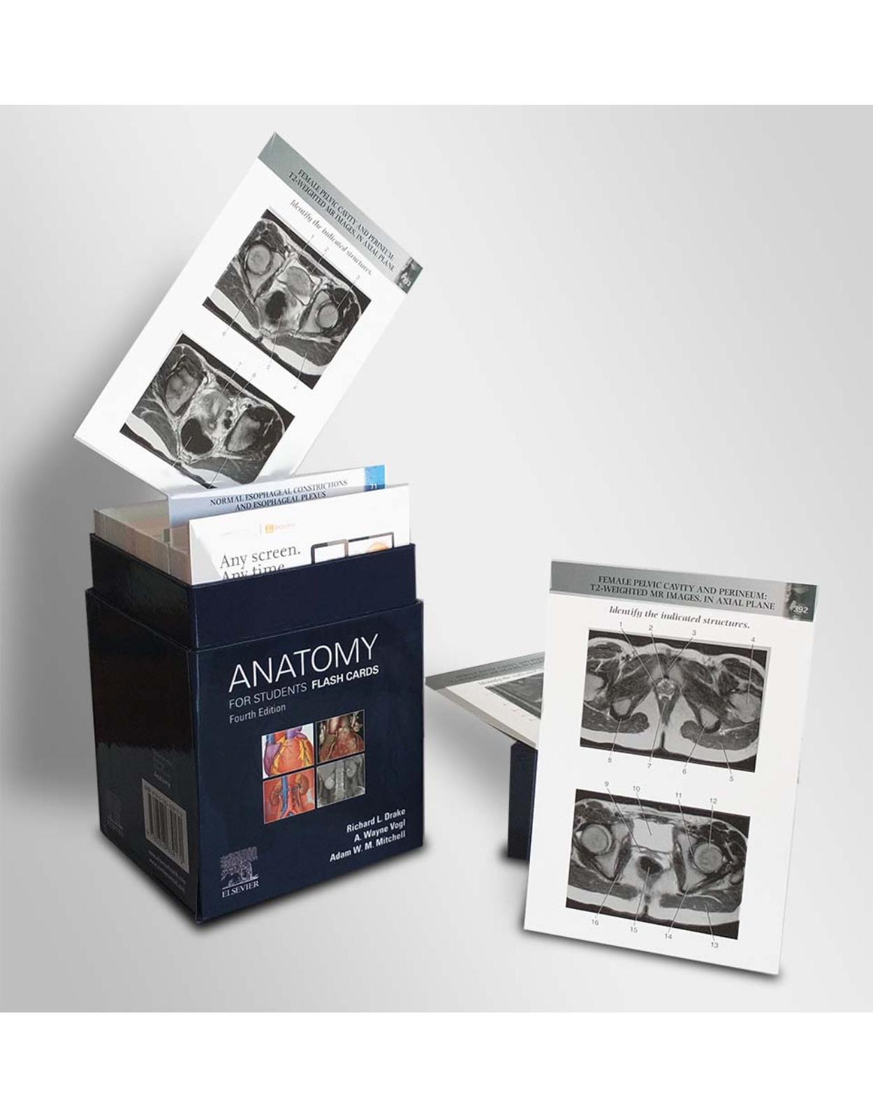
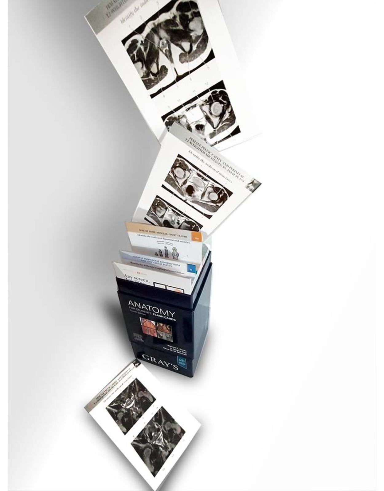
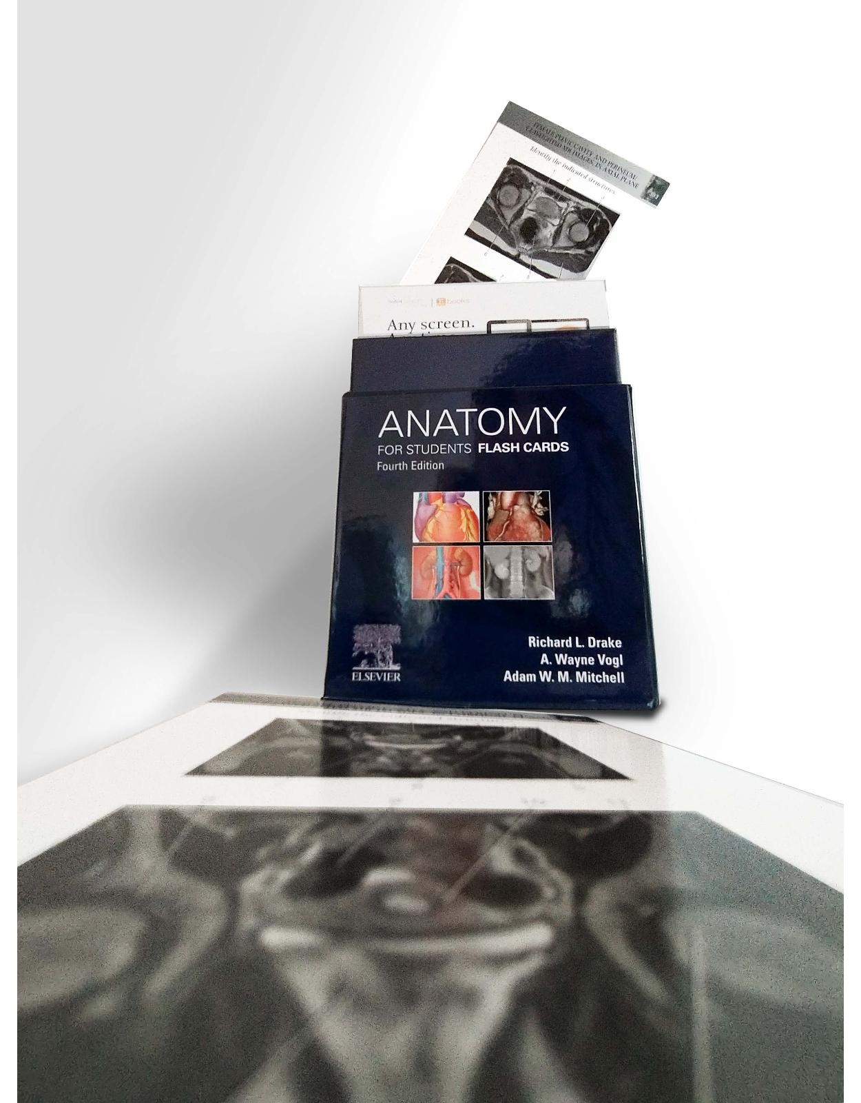
Gray’s Anatomy for Students Flash Cards
Produs indisponibil momentan. Pentru comenzi va rugam trimiteti mail la adresa depozit2@prior.ro sau contactati-ne la numarul de telefon 021 210 89 28 Vedeti mai jos alte produse similare disponibile.
Disponibilitate: Acest produs nu este momentan in stoc
Editura: Elsevier
Limba: Engleza
Nr. pagini: 820 cards
Coperta: Flash card
Dimensiuni: 12.1 x 10.2 x 17.2 cm
An aparitie: 28 May 2019
Description:
Study efficiently while being confident in your mastery of the most important anatomical concepts! Flashcards have been thoroughly revised to reflect the updates made to the companion text, Gray's Anatomy for Students, 4th Edition.
Understand the clinical relevance of your anatomical knowledge with clinical imaging cards.
Conveniently access all of the need-to-know anatomy information! Each card presents beautiful 4-color artwork or a radiologic image of a particular structure/area of the body, with numbered leader lines indicating anatomical structures; labels to the structures are listed by number on the reverse, in addition to relevant functions, clinical correlations, and more.
Fully grasp the practical applications of anatomy with "In the Clinic" discussions on most cards, which relate structures to corresponding clinical disorders; a page reference to the companion textbook (Gray's Anatomy for Students, 4th Edition) facilitates access to further information.
Access a clear, visual review of key concepts with wiring diagrams that detail the innervation of nerves to organs and other body parts, as well as muscle cards covering functions and attachments.
Table of Contents:
Section 1 Overview
1 Surface Anatomy: Male Anterior View
2 Surface Anatomy: Female Posterior View
3 Skeleton: Anterior View
4 Skeleton: Posterior View
5 Muscles: Anterior View
6 Muscles: Posterior View
7 Vascular System: Arteries
8 Vascular System: Veins
Section 2 Back
9 Skeletal Framework: Vertebral Column
10 Skeletal Framework: Typical Vertebra
11 Skeletal Framework: Vertebra 1
12 Skeletal Framework: Atlas, Axis, and Ligaments
13 Skeletal Framework: Vertebra 2
14 Skeletal Framework: Vertebra 3
15 Skeletal Framework: Sacrum and Coccyx
16 Skeletal Framework: Vertebra Radiograph I
17 Skeletal Framework: Vertebra Radiograph II
18 Skeletal Framework: Vertebra Radiograph III
19 Skeletal Framework: Intervertebral Joints
20 Skeletal Framework: Intervertebral Foramen
21 Skeletal Framework: Vertebral Ligaments
22 Skeletal Framework: Intervertebral Disc Protrusion
23 Muscles: Superficial Group
24 Muscles: Trapezius Innervation and Blood Supply
25 Muscles: Intermediate Group
26 Muscles: Erector Spinae
27 Muscles: Transversospinalis and Segmentals
28 Muscles: Suboccipital Region
29 Spinal Cord
30 Spinal Cord Details
31 Spinal Nerves
32 Spinal Cord Arteries
33 Spinal Cord Arteries Detail
34 Spinal Cord Meninges
Section 3 Thorax
35 Thoracic Skeleton
36 Typical Rib
37 Rib I Superior Surface
38 Sternum
39 Vertebra, Ribs, and Sternum
40 Thoracic Wall
41 Thoracic Cavity
42 Intercostal Space with Nerves and Vessels
43 Pleural Cavity
44 Pleura
45 Parietal Pleura
46 Right Lung
47 Left Lung
48 CT: Left Pulmonary Artery
49 CT: Right Pulmonary Artery
50 Mediastinum: Subdivisions
51 Pericardium
52 Pericardial Sinuses
53 Anterior Surface of the Heart
54 Diaphragmatic Surface and Base of the Heart
55 Right Atrium
56 Right Ventricle
57 Left Atrium
58 Left Ventricle
59 Plain Chest Radiograph
60 MRI: Chambers of the Heart
61 Coronary Arteries
62 Coronary Veins
63 Conduction System
64 Superior Mediastinum
65 Superior Mediastinum: Cross Section
66 Superior Mediastinum: Great Vessels
67 Superior Mediastinum: Trachea and Esophagus
68 Mediastinum: Right Lateral View
69 Mediastinum: Left Lateral View
70 Posterior Mediastinum
71 Normal Esophageal Constrictions and Esophageal Plexus
72 Thoracic Aorta and Branches
73 Azygos System of Veins and Thoracic Duct
74 Thoracic Sympathetic Trunks and Splanchnic Nerves
Section 4 Abdomen
75 Abdominal Wall: Nine-Region Pattern
76 Abdominal Wall: Layers Overview
77 Abdominal Wall: Transverse Section
78 Rectus Abdominis
79 Rectus Sheath
80 Inguinal Canal
81 Spermatic Cord
82 Round Ligament of the Uterus
83 Inguinal Region: Internal View
84 Viscera: Anterior View
85 Viscera: Anterior View, Small Bowel Removed
86 Stomach
87 Double-Contrast Radiograph: Stomach and Duodenum
88 Duodenum
89 Radiograph: Jejunum and Ileum
90 Large Intestine
91 Barium Radiograph: Large Intestine
92 Liver
93 CT: Liver
94 Pancreas
95 CT: Pancreas
96 Bile Drainage
97 Arteries: Arterial Supply of Viscera
98 Arteries: Celiac Trunk
99 Arteries: Superior Mesenteric
100 Arteries: Inferior Mesenteric
101 Veins: Portal System
102 Viscera: Innervation
103 Posterior Abdominal Region: Overview
104 Posterior Abdominal Region: Bones
105 Posterior Abdominal Region: Muscles
106 Diaphragm
107 Anterior Relationships of Kidneys
108 Internal Structure of the Kidney
109 CT: Renal Pelvis
110 Renal and Suprarenal Gland Vessels
111 Abdominal Aorta
112 Inferior Vena Cava
113 Urogram: Pathway of Ureter
114 Lumbar Plexus
Section 5 Pelvis and Perineum
115 Pelvis
116 Pelvic Bone
117 Ligaments
118 Muscles: Pelvic Diaphragm and Lateral Wall
119 Perineal Membrane and Deep Perineal Pouch
120 Viscera: Female Overview
121 Viscera: Male Overview
122 Male Reproductive System
123 Female Reproductive System
124 Uterus and Uterine Tubes
125 Sacral Plexus
126 Internal Iliac Posterior Trunk
127 Internal Iliac Anterior Trunk
128 Female Perineum
129 Male Perineum
130 Anal Triangle Cross Section
131 Superficial Perineal Pouch: Muscles
132 MRI: Male Pelvic Cavity and Perineum
133 Deep Perineal Pouch: Muscles
134 MRI: Female Pelvic Cavity and Perineum
Section 6 Lower Limb
135 Skeleton: Overview
136 Acetabulum
137 Femur
138 Hip Joint Ligaments
139 Ligament of Head of Femur
140 Radiograph: Hip Joint
141 CT: Hip Joint
142 Femoral Triangle
143 Saphenous Vein
144 Anterior Compartment: Muscles
145 Anterior Compartment: Muscle Attachments
146 Femoral Artery
147 Medial Compartment: Muscles
148 Medial Compartment: Muscle Attachments
149 Obturator Nerve
150 Gluteal Region: Muscles
151 Gluteal Region: Muscle Attachments I
152 Gluteal Region: Muscle Attachments II
153 Gluteal Region: Arteries
154 Gluteal Region: Nerves
155 Sacral Plexus
156 Posterior Compartment: Muscles
157 Posterior Compartment: Muscle Attachments
158 Sciatic Nerve
159 Knee: Anterolateral View
160 Knee: Menisci and Ligaments
161 Knee: Collateral Ligaments
162 MRI: knee joint
163 Radiographs: Knee Joint
164 Knee: Popliteal Fossa
165 Leg: Bones
166 Leg Posterior Compartment: Muscles
167 Leg Posterior Compartment: Muscle Attachments I
168 Leg Posterior Compartment: Muscle Attachments II
169 Leg Posterior Compartment: Arteries and Nerves
170 Leg Lateral Compartment: Muscles
171 Leg Lateral Compartment: Muscle Attachments
172 Leg Lateral Compartment: Nerves
173 Leg Anterior Compartment: Muscles
174 Leg Anterior Compartment: Muscle Attachments
175 Leg Anterior Compartment: Arteries and Nerves
176 Foot: Bones
177 Radiograph: Foot
178 Foot: Ligaments
179 Radiograph: Ankle
180 Dorsal Foot: Muscles
181 Dorsal Foot: Muscle Attachments
182 Dorsal Foot: Arteries
183 Dorsal Foot: Nerves
184 Tarsal Tunnel
185 Sole of Foot: Muscles, First Layer
186 Sole of Foot: Muscles, Second Layer
187 Sole of Foot: Muscles, Third Layer
188 Sole of Foot: Muscles, Fourth Layer
189 Sole of Foot: Muscle Attachments, First and Second Layers
190 Sole of Foot: Muscle Attachments, Third Layer
191 Sole of Foot: Arteries
192 Sole of Foot: Nerves
Section 7 Upper Limb
193 Overview: Skeleton
194 Clavicle
195 Scapula
196 Humerus
197 Sternoclavicular and Acromioclavicular Joints
198 Multidetector CT: Sternoclavicular Joint
199 Radiograph: Acromioclavicular Joint
200 Shoulder Joint
201 Radiograph: Glenohumeral Joint
202 Pectoral Region: Breast
203 Pectoralis Major
204 Pectoralis Minor: Nerves and Vessels
205 Posterior Scapular Region: Muscles
206 Posterior Scapular Region: Muscle Attachments
207 Posterior Scapular Region: Arteries and Nerves
208 Axilla: Vessels
209 Axilla: Arteries
210 Axilla: Nerves
211 Axilla: Brachial Plexus
212 Axilla: Lymphatics
213 Humerus: Posterior View
214 Distal Humerus
215 Proximal End of Radius and Ulna
216 Arm Anterior Compartment: Biceps
217 Arm Anterior Compartment: Muscles
218 Arm Anterior Compartment: Muscle Attachments
219 Arm Anterior Compartment: Arteries
220 Arm Anterior Compartment: Veins
221 Arm Anterior Compartment: Nerves
222 Arm Posterior Compartment: Muscles
223 Arm Posterior Compartment: Muscle Attachments
224 Arm Posterior Compartment: Nerves and Vessels
225 Elbow Joint
226 Cubital Fossa
227 Radius
228 Ulna
229 Radiographs: Elbow Joint
230 Radiograph: Forearm
231 Wrist and Bones of Hand
232 Radiograph: Wrist
233 Radiographs: Hand and Wrist Joint
234 Forearm Anterior Compartment: Muscles, First Layer
235 Forearm Anterior Compartment: Muscle Attachments, Superficial Layer
236 Forearm Anterior Compartment: Muscles, Second Layer
237 Forearm Anterior Compartment: Muscles, Third Layer
238 Forearm Anterior Compartment: Muscle Attachments, Intermediate and Deep Layers
239 Forearm Anterior Compartment: Arteries
240 Forearm Anterior Compartment: Nerves
241 Forearm Posterior Compartment: Muscles, Superficial Layer
242 Forearm Posterior Compartment: Muscle Attachments, Superficial Layer
243 Forearm Posterior Compartment: Outcropping Muscles
244 Forearm Posterior Compartment: Muscle Attachments, Deep Layer
245 Forearm Posterior Compartment: Nerves and Arteries
246 Hand: Cross Section Through Wrist
247 Hand: Superficial Palm
248 Hand: Thenar and Hypothenar Muscles
249 Palm of Hand: Muscle Attachments, Thenar and Hypothenar Muscles
250 Lumbricals
251 Adductor Muscles
252 Interosseous Muscles
253 Palm of Hand: Muscle Attachments
254 Superficial Palmar Arch
255 Deep Palmar Arch
256 Median Nerve
257 Ulnar Nerve
258 Radial Nerve
259 Dorsal Venous Arch
Section 8 Head and Neck
260 Skull: Anterior View
261 Multidetector CT: Anterior View of Skull
262 Skull: Lateral View
263 Multidetector CT: Lateral View of Skull
264 Skull: Posterior View
265 Skull: Superior View
266 Skull: Inferior View
267 Skull: Anterior Cranial Fossa
268 Skull: Middle Cranial Fossa
269 Skull: Posterior Cranial Fossa
270 Meninges
271 Dural Septa
272 Meningeal Arteries
273 Blood Supply to Brain
274 Magnetic Resonance Angiogram: Carotid and Vertebral Arteries
275 Circle of Willis
276 Dural Venous Sinuses
277 Cavernous Sinus
278 Cavernous Sinus
279 Cranial Nerves: Floor of Cranial Cavity
280 Facial Muscles
281 Lateral Face
282 Sensory Nerves of the Head
283 Vessels of the Lateral Face
284 Scalp
285 Orbit: Bones
286 Lacrimal Apparatus
287 Orbit: Extra-ocular Muscles
288 MRI: Muscles of the Eyeball
289 Superior Orbital Fissure and Optic Canal
290 Orbit: Superficial Nerves
291 Orbit: Deep Nerves
292 Eyeball
293 Visceral Efferent (Motor) Innervation: Lacrimal Gland
294 Visceral Efferent (Motor) Innervation: Eyeball (Iris and Ciliary Body)
295 Visceral Efferent (Motor) Pathways Through Pterygopalatine Fossa
296 External Ear
297 External, Middle, and Internal Ear
298 Tympanic Membrane
299 Middle Ear: Schematic View
300 Internal Ear
301 Infratemporal Region: Muscles of Mastication
302 Infratemporal Region: Muscles
303 Infratemporal Region: Arteries
304 Infratemporal Region: Nerves, Part 1
305 Infratemporal Region: Nerves, Part 2
306 Parasympathetic Innervation of Salivary Glands
307 Pterygopalatine Fossa: Gateways
308 Pterygopalatine Fossa: Nerves
309 Pharynx: Posterior View of Muscles
310 Pharynx: Lateral View of Muscles
311 Pharynx: Midsagittal Section
312 Pharynx: Posterior View, Opened
313 Larynx: Overview
314 Larynx: Cartilage and Ligaments
315 Larynx: Superior View of Vocal Ligaments
316 Larynx: Posterior View
317 Larynx: Laryngoscopic Images
318 Larynx: Intrinsic Muscles
319 Larynx: Nerves
320 Nasal Cavity: Paranasal Sinuses
321 Radiographs: Nasal Cavities and Paranasal Sinuses
322 CT: Nasal Cavities and Paranasal Sinuses
323 Nasal Cavity: Nasal Septum
324 Nasal Cavity: Lateral Wall, Bones
325 Nasal Cavity: Lateral Wall, Mucosa, and Openings
326 Nasal Cavity: Arteries
327 Nasal Cavity: Nerves
328 Oral Cavity: Overview
329 Oral Cavity: Floor
330 Oral Cavity: Tongue
331 Oral Cavity: Sublingual Glands
332 Oral Cavity: Glands
333 Oral Cavity: Salivary Gland Nerves
334 Oral Cavity: Soft Palate (Overview)
335 Oral Cavity: Palate, Arteries, and Nerves
336 Oral Cavity: Teeth
337 Neck: Triangles
338 Neck: Fascia
339 Neck: Superficial Veins
340 Neck: Anterior Triangle, Infrahyoid Muscles
341 Neck: Anterior Triangle, Carotid System
342 Neck: Anterior Triangle, Glossopharyngeal Nerve
343 Neck: Anterior Triangle, Vagus Nerve
344 Neck: Anterior Triangle, Hypoglossal Nerve, and Ansa Cervicalis
345 Neck: Anterior Triangle, Anterior View Thyroid
346 Neck: Anterior Triangle, Posterior View Thyroid
347 Neck: Posterior Triangle, Muscles
348 Neck: Posterior Triangle, Nerves
349 Base of Neck
350 Base of Neck: Arteries
351 Base of Neck: Lymphatics
Section 9 Surface Anatomy
352 Back Surface Anatomy
353 End of Spinal Cord: Lumbar Puncture
354 Thoracic Skeletal Landmarks
355 Heart Valve Auscultation
356 Lung Auscultation 1
357 Lung Auscultation 2
358 Referred Pain: Heart
359 Inguinal Hernia I
360 Inguinal Hernia II
361 Inguinal Hernia III
362 Referred Abdominal Pain
363 Female Perineum
364 Male Perineum
365 Gluteal Injection Site
366 Femoral Triangle Surface Anatomy
367 Popliteal Fossa
368 Tarsal Tunnel
369 Lower Limb Pulse Points
370 Upper Limb Pulse Points
371 Head and Neck Pulse Points
Section 10 Nervous System
372 Brain: Base of Brain Cranial Nerves
373 Spinal Cord
374 Spinal Nerve
375 Heart Sympathetics
376 Gastrointestinal Sympathetics
377 Parasympathetics
378 Parasympathetic Ganglia
379 Pelvic Autonomics
Section 11 Imaging
380 Mediastinum: CT Images, Axial Plane
381 Mediastinum: CT Images, Axial Plane
382 Mediastinum: CT Images, Axial Plane
383 Stomach and Duodenum: Double-Contrast Radiograph
384 Jejunum and Ileum: Radiograph
385 Large Intestine: Radiograph, Using Barium
386 Liver: Abdominal CT Scan with Contrast, in Axial Plane
387 Pancreas: Abdominal CT Scan with Contrast, in Axial Plane
388 Male Pelvic Cavity and Perineum: T2-Weighted MR Images, in Axial Plane
389 Male Pelvic Cavity and Perineum: T2-Weighted MR Images, in Axial Plane
390 Female Pelvic Cavity and Perineum: T2-Weighted MR Images, in Sagittal Plane
391 Female Pelvic Cavity and Perineum: T2-Weighted MR Images, in Coronal Plane
392 Female Pelvic Cavity and Perineum: T2-Weighted MR Images, in Axial Plane
393 Female Pelvic Cavity and Perineum: T2-Weighted MR Images, in Axial Plane
| An aparitie | 28 May 2019 |
| Autor | Richard Drake PhD FAAA , A. Wayne Vogl PhD FAAA , Adam W. M. Mitchell MB BS FRCS FRCR |
| Dimensiuni | 12.1 x 10.2 x 17.2 cm |
| Editura | Elsevier |
| Format | Flash card |
| ISBN | 9780323639170 |
| Limba | Engleza |
| Nr pag | 820 cards |
-
 72100 lei 68700 lei
 -

-

-

-
-


Clientii ebookshop.ro nu au adaugat inca opinii pentru acest produs. Fii primul care adauga o parere, folosind formularul de mai jos.