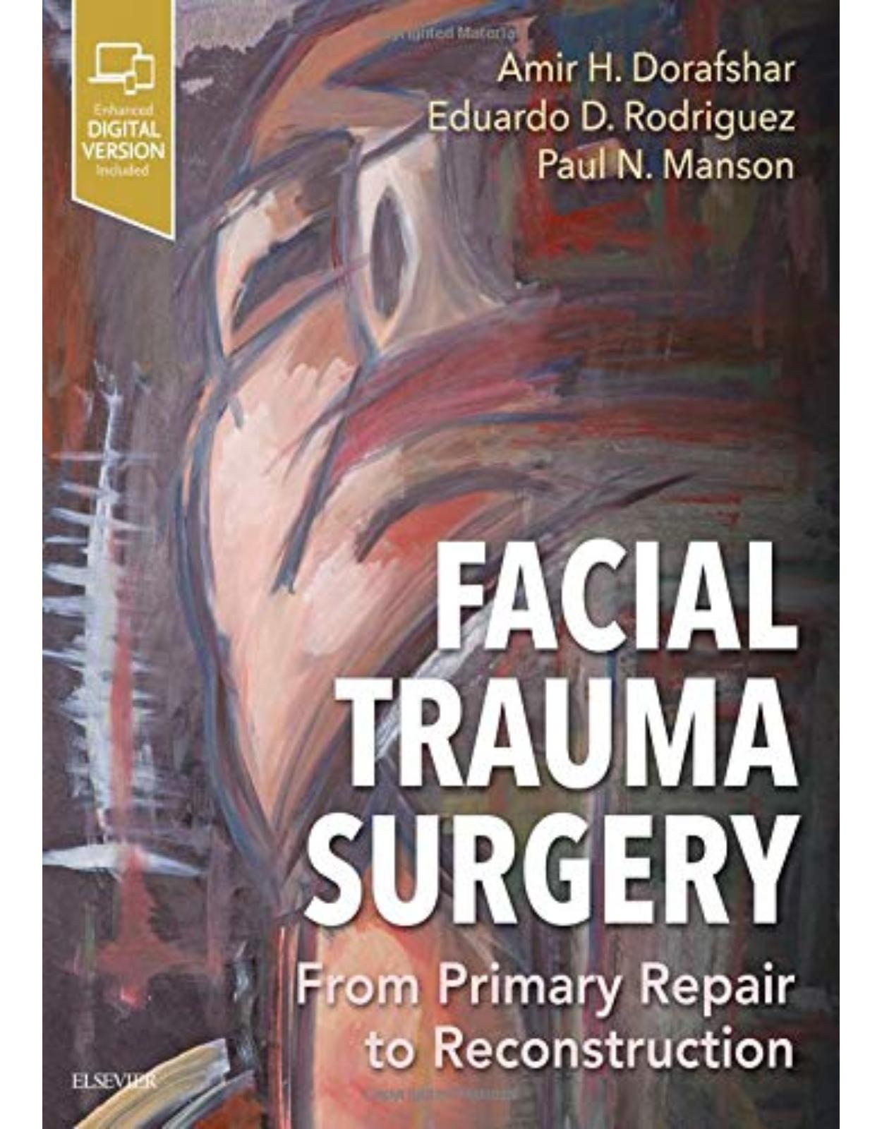
Facial Trauma Surgery: From Primary Repair to Reconstruction Hardcover
Livrare gratis la comenzi peste 500 RON. Pentru celelalte comenzi livrarea este 20 RON.
Disponibilitate: La comanda in aproximativ 4-6 saptamani
Editura: Elsevier
Limba: Engleza
Nr. pagini: 560
Coperta: Hardcover
Dimensiuni: 21.6 x 2.5 x 27.9 cm
An aparitie: 2 May 2019
Description:
Offering authoritative guidance and a multitude of high-quality images, Facial Trauma Surgery: From Primary Repair to Reconstruction is the first comprehensive textbook of its kind on treating primary facial trauma and delayed reconstruction of both the soft tissues and craniofacial bony skeleton. This unique volume is a practical, complete reference for clinical presentation, fracture pattern, classification, and management of patients with traumatic facial injury, helping you provide the best possible outcomes for patients’ successful reintegration into work and society.
- Explains the basic principles and concepts of primary traumatic facial injury repair and secondary facial reconstruction.
- Offers expert, up-to-date guidance from global leaders in plastic and reconstructive surgery, otolaryngology and facial plastic surgery, oral maxillofacial surgery, neurosurgery, and oculoplastic surgery.
- Covers innovative topics such as virtual surgical planning, 3D printing, intraoperative surgical navigation, post-traumatic injury, treatment of facial pain, and the roles of microsurgery and facial transplantation in the treatment facial traumatic injuries.
- Includes an end commentary in every chapter provided by Dr. Paul Manson, former Chief of Plastic Surgery at Johns Hopkins Hospital and a pioneer in the field of acute treatment of traumatic facial injuries.
- Features superb photographs and illustrations throughout, as well as evidence-based summaries in current areas of controversy.
1. Table of Contents
2. Copyright
3. Video Contents
4. Foreword
5. “Precision Medicine” and Facial Injuries, 2018
6. References
7. Preface
8. List of Contributors
9. Acknowledgments
10. Dedication
11. Section 1 Primary Injury
12. 1.1 Assessment of the Patient With Traumatic Facial Injury
13. Background
14. Surgical Anatomy
15. Classification
16. Clinical Presentation
17. Radiological Evaluation
18. Surgical Treatments
19. References
20. References
21. 1.2 Radiological Evaluation of the Craniofacial Skeleton
22. Background
23. Surgical Anatomy
24. Radiological Evaluation
25. Classification
26. Postoperative Imaging
27. Acute and Long-Term Complications
28. Mandible
29. References
30. References
31. 1.3 Intraoperative Imaging and Postoperative Quality Control
32. Background
33. Surgical Anatomy in 3D Imaging
34. Radiological Evaluation
35. Surgical Techniques
36. Postoperative Course
37. Acute Complications
38. Long-Term Complications
39. The Need for Quality Control
40. References
41. 1.4 Primary Repair of Soft Tissue Injury and Soft Tissue Defects
42. Background
43. Surgical Anatomy
44. Clinical Presentation
45. Radiological Evaluation
46. Classification of Soft Tissue Injuries
47. Surgical Techniques
48. References
49. References
50. 1.5 Traumatic Facial Nerve Injury
51. Introduction
52. Epidemiology and Etiology
53. Surgical Anatomy
54. History and Physical Exam
55. Location, Mechanism, and Duration of Injury – Effects on Management Strategies
56. Additional Procedures
57. Complications
58. Conclusion
59. References
60. 1.6 Diagnosis and Multimodality Management of Skull Base Fractures and Cerebrospinal Fluid Leaks
61. Background
62. Surgical Anatomy
63. Clinical Presentation
64. Radiological Evaluation
65. Considerations in the Initial Management of Skull Base CSF Leaks
66. Surgical Techniques
67. Early and Delayed Complications
68. Case Example
69. Conclusions
70. References
71. 1.7 Frontal Bone and Frontal Sinus Injuries
72. Background
73. Anatomy
74. Etiology and Incidence
75. History and Physical Examination
76. Radiography
77. Surgical Approaches
78. Treatment Strategies
79. Treatment Algorithm
80. Complications and Long-Term Follow Up
81. References
82. References
83. 1.8 Endoscopic Approaches to Frontal and Maxillary Sinus Fractures
84. Background
85. Surgical Anatomy
86. Clinical Presentation
87. Radiological Evaluation
88. Surgical Indications
89. Transnasal Endoscopic Approach to Frontal Sinus Fractures
90. Endoscopic Approaches to Repair of Orbital Floor Fractures
91. Postoperative Course
92. Complications
93. Conclusion
94. References
95. 1.9 Orbital Fractures
96. Background
97. Surgical Anatomy
98. Clinical Presentation
99. Radiological Evaluation
100. Classification
101. Surgical Indications
102. Surgical Techniques
103. Postoperative Course
104. Acute Complications
105. Long-Term Complications
106. References
107. 1.10 Nasal Fractures
108. Background
109. Surgical Anatomy
110. Clinical Presentation
111. Radiological Evaluation
112. Classification
113. Surgical Indications
114. Surgical Techniques
115. Postoperative Course
116. Complications
117. References
118. 1.11 Naso-orbito-ethmoid (NOE) Fractures
119. Background
120. Surgical Anatomy
121. Clinical Presentation
122. Radiological Evaluation
123. Classification
124. Surgical Indications
125. Surgical Techniques
126. Postoperative Course
127. Acute Complications
128. Long-Term Complications
129. References
130. References
131. 1.12 Orbitozygomaticomaxillary Complex Fractures
132. Background
133. Incidence and Etiology
134. Surgical Anatomy
135. Clinical Presentation
136. Radiological Evaluation
137. Classification
138. Surgical Indications
139. Surgical Techniques
140. Postoperative Course
141. Acute Complications
142. Long-Term Complications
143. Conclusion
144. References
145. References
146. 1.13 Le Fort Fractures
147. Background
148. Incidence
149. Etiology
150. Surgical Anatomy
151. Clinical Presentation
152. Radiological Evaluation
153. Classification
154. Surgical Indications
155. Surgical Goals/Techniques
156. Postoperative Course
157. Acute Complications
158. Long-Term Complications
159. References
160. References
161. 1.14 Mandible Fractures
162. Background
163. Epidemiology
164. Surgical Anatomy
165. Classification
166. Clinical Presentation
167. Physical Examination
168. Radiological Examination
169. Surgical Indications
170. Surgical Approaches/Techniques
171. Postoperative Course
172. Complications
173. References
174. 1.15 Fractures of the Condylar Process of the Mandible
175. Background
176. Surgical Anatomy and Clinical Presentation
177. Radiological Evaluation
178. Terminology
179. Classification of Condylar Process Fractures
180. Treatment of Condylar Process Fractures
181. Trauma-Derived Temporomandibular Disorders
182. References
183. References
184. 1.16 Complications of Mandibular Fractures
185. The Problem
186. Nature of Complications
187. Evolution of Complications
188. Incidence of Complications
189. Etiologic Factors in Mandibular Fracture Complications
190. Infection, Delayed, Non- and Malunion, Hardware Exposure, Hardware Failure
191. Paresthesia/Paresis
192. Complications of Condylar Fractures
193. Patient Noncompliance
194. Conclusion
195. References
196. 1.17 Temporal Bone Fractures
197. Background
198. Etiology
199. Temporal Bone Anatomy
200. Clinical Presentation
201. Classification of Temporal Bone Fractures
202. Management, Surgical Indications, and Techniques
203. Long-Term Complications
204. Conclusion
205. Reference
206. References
207. 1.18 Dentoalveolar Trauma
208. Background
209. Surgical Anatomy
210. Clinical Presentation
211. Radiological Evaluation
212. Classification
213. Management
214. Surgical Techniques
215. Acute Complications
216. Long-Term Complications
217. References
218. References
219. 1.19 Management of Panfacial Fractures
220. Background
221. Surgical Anatomy
222. Clinical Presentation
223. Radiological Evaluation
224. Presurgical Preparation
225. Surgical Techniques
226. Postoperative Course
227. Acute Complications
228. Long-Term Complications
229. Case Examples
230. References
231. References
232. 1.20 Characteristics of Ballistic and Blast Injuries
233. Introduction
234. Background
235. Components of Ballistic Missiles
236. Components of Improvised Explosive Devices
237. The Principles of Velocity
238. Characteristics of Ballistic Injuries
239. Conclusion
240. Key Points
241. Bibliography
242. 1.21 Geriatric and Edentulous Maxillary and Mandibular Fractures
243. Diagnosis
244. Mandible Fractures
245. Complications
246. Maxillary Fractures
247. Complications
248. References
249. References
250. Section 2 Pediatric Facial Injury
251. 2.1 Pediatric Skull Fractures
252. Background
253. Epidemiology
254. Pediatric Skull and Fracture Anatomy
255. Clinical Presentation
256. Radiological Evaluation
257. Surgical Indications
258. Surgical Techniques
259. Postoperative Course
260. Illustrative Case Examples
261. Complications
262. Conclusion
263. References
264. 2.2 Superior Pediatric Orbital and Frontal Skull Fractures
265. Background and Incidence
266. Surgical Anatomy
267. Clinical Presentation
268. Radiological Evaluation
269. Surgical Indications
270. Surgical Techniques
271. Acute Complications
272. Long-Term Complications
273. References
274. 2.3 Pediatric Orbital Fractures
275. Background
276. Surgical Anatomy
277. Classification
278. Clinical Presentation
279. Radiographic Evaluation
280. Surgical Indications
281. Surgical Technique
282. Postoperative Course
283. Acute Complications
284. Long-Term Complications
285. Conclusions
286. References
287. 2.4 Pediatric Midface Fractures
288. Background
289. Surgical Anatomy
290. Clinical Presentation
291. Radiological Evaluation
292. Classification
293. Surgical Indications
294. Maxillomandibular Fixation
295. Surgical Techniques
296. Postoperative Course
297. Conclusion
298. Reference
299. References
300. 2.5 Pediatric Mandible Fractures
301. Background
302. Embryology and Growth of the Mandible
303. Anatomy of the Mandible
304. Stages of Dentition
305. Fracture Patterns in Pediatric Populations
306. Evaluation
307. Treatment
308. Major Complications and Management
309. Conclusion
310. References
311. Section 3 Secondary Reconstruction and Restoration
312. 3.1 Reconstruction of Full-Thickness Frontocranial Defects
313. Background
314. Case Examples
315. Conclusions
316. References
317. References
318. 3.2 Pediatric Cranial Reconstruction
319. Background
320. Clinical Presentation
321. Pediatric Cranioplasty – Surgical Techniques
322. References
323. 3.3 Secondary Reconstruction of Facial Soft Tissue Injury and Defects
324. Background and Etiology
325. Initial Assessment
326. Principles of Secondary Soft Tissue Correction
327. Soft Tissue Contour Augmentation
328. Temporal Contour Deformity
329. Cheek Ptosis
330. Periorbital Deformity
331. Nasal Deformities
332. Brow Ptosis
333. Scar Revision
334. Resurfacing Techniques
335. Traumatic Tattooing
336. Excisional Techniques
337. Tissue Expansion
338. References
339. References
340. 3.4 Ocular Considerations
341. Background
342. Surgical Anatomy
343. Clinical Assessment
344. Entropion
345. Ectropion
346. Blink
347. Ptosis
348. Epiphora
349. Surgical Techniques
350. Postoperative Considerations
351. Long-Term Complications
352. References
353. 3.5 Secondary Nasoethmoid Fracture Repair
354. Background
355. Surgical Anatomy
356. Clinical Presentation
357. Radiological Evaluation
358. Classification (Fig. 3.5.9)
359. Surgical Indications
360. Surgical Techniques (Fig. 3.5.11)
361. Postoperative Course
362. Complications
363. Conclusion
364. References
365. References
366. 3.6 Posttraumatic Nasal Deformities
367. Background
368. Surgical Anatomy
369. Clinical Presentation
370. Surgical Techniques
371. Case Examples
372. Postoperative Course
373. Complications
374. Conclusion
375. References
376. 3.7 Secondary Orbital Reconstruction
377. Background
378. Clinical Presentation
379. Radiological Evaluation
380. Surgical Indications
381. Surgical Techniques
382. Postoperative Course
383. Complications
384. References
385. 3.8 Secondary Midfacial Reconstruction
386. Background
387. Central Midface and Medial Canthal Deformity
388. Zygoma Fractures Secondary Deformity
389. Le Fort Fractures Secondary Deformity
390. Conclusions
391. References
392. 3.9 Secondary Osteotomies of the Maxilla and Mandible, and Management of Occlusion
393. Background
394. Patient Evaluation
395. Case Examples
396. Conclusions
397. References
398. 3.10 Secondary Traumatic TMJ Reconstruction
399. Background
400. Anatomy of the TMJ
401. Posttraumatic Indications for TMJ Reconstruction
402. Autogenous Graft Failure
403. Imaging of the TMJ
404. TMJ Reconstructive Techniques
405. Surgical Technique (Stock Prosthesis)
406. Complications
407. References
408. 3.11 Maxillofacial Prosthodontics
409. Background
410. Oral and Dental Anatomy Evaluation
411. Intraoral Prosthetic Rehabilitation
412. Soft Palate Evaluation
413. Tongue Evaluation
414. Extraoral Evaluation
415. Osseointegrated Dental Implants
416. Conclusion
417. References
418. 3.12 Custom Craniofacial Implants
419. Background
420. Clinical Presentation
421. Radiological Evaluation
422. CAD/CAM Facial Implants
423. Preferred Surgical Techniques
424. Postoperative Course
425. Reference
426. References
427. 3.13 Secondary Microvascular Reconstruction of the Traumatic Facial Injury
428. Background
429. Surgical Anatomy
430. Clinical Presentation
431. Radiologic Evaluation
432. Surgical Indications
433. Surgical Technique
434. Postoperative Course
435. Complications
436. Case Examples
437. References
438. References
439. 3.14 Virtual Surgical Planning
440. Background
441. Contemporary Uses of CAS
442. Primary Reconstruction
443. Secondary Reconstruction
444. Future of CAS in Craniofacial Surgery
445. References
446. 3.15 Posttraumatic Facial Pain
447. Background
448. Differential Diagnosis of Posttraumatic Facial Pain
449. Surgical Treatment of A Painful Neuroma (Box 3.15.2)
450. Frontal Branch of the Trigeminal Nerve (V1)
451. Cervical Plexus Injury and Facial Pain
452. Occipital Nerves and Facial Pain
453. Reference
454. References
455. 3.16 Secondary Nerve Reconstruction
456. Background
457. Trigeminal Anatomy
458. Peripheral Nerve Injury and Histopathology
459. Clinical Presentation
460. Surgical Techniques
461. Follow-up
462. References
463. 3.17 Facial Transplantation
464. Background
465. Surgical Indications
466. Classification
467. Surgical Techniques
468. Postoperative Course
469. Acute Complications
470. Long-Term Complications
471. References
472. Appendix 1 Evidence-Based Medicine
| An aparitie | 2 May 2019 |
| Autor | Amir H Dorafshar MBChB FACS FAAP |
| Dimensiuni | 21.6 x 2.5 x 27.9 cm |
| Editura | Elsevier |
| Format | Hardcover |
| ISBN | 9780323497558 |
| Limba | Engleza |
| Nr pag | 560 |
-
1,32700 lei 1,20500 lei
-
2,07800 lei 1,76500 lei






Clientii ebookshop.ro nu au adaugat inca opinii pentru acest produs. Fii primul care adauga o parere, folosind formularul de mai jos.