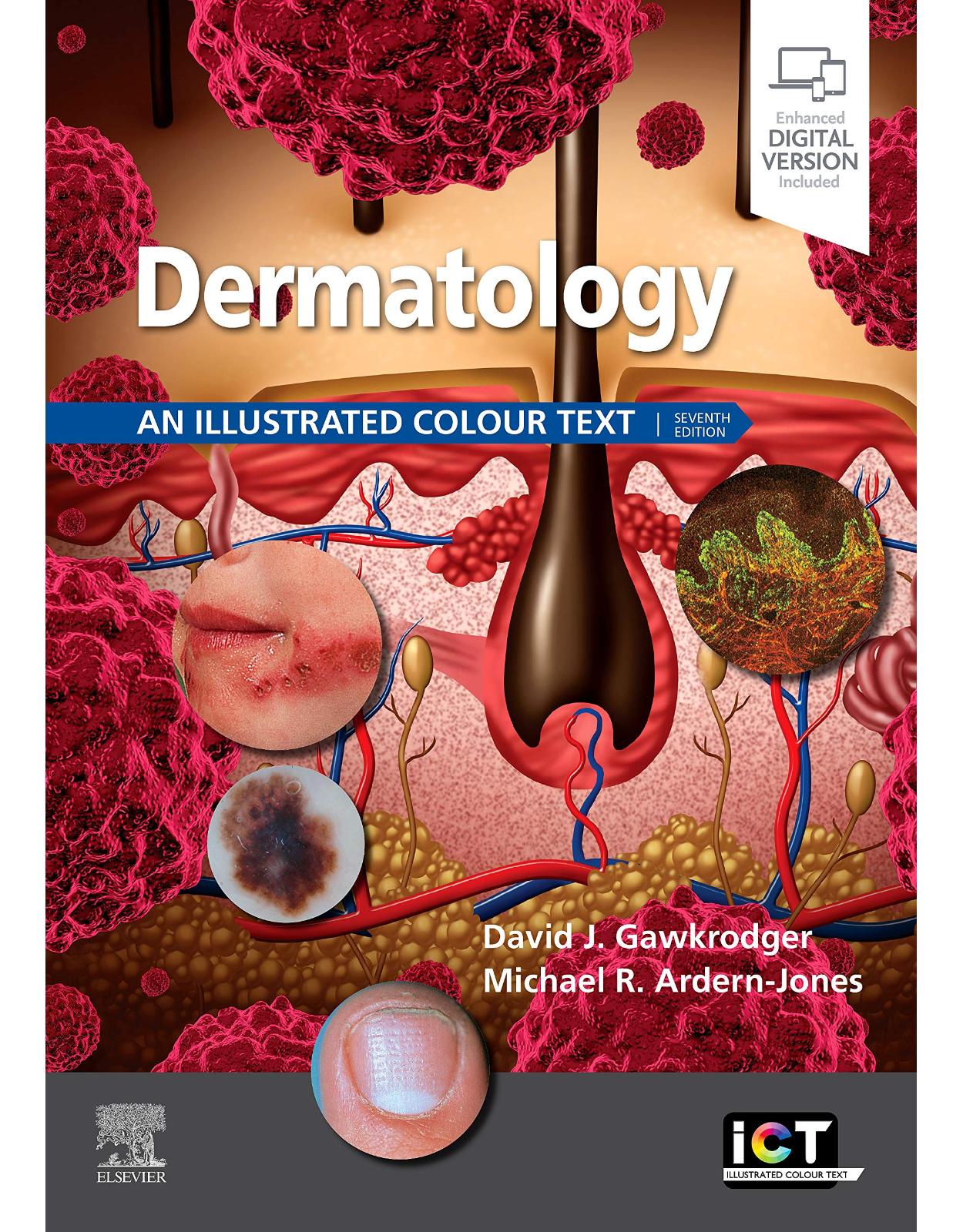
Dermatology: An Illustrated Colour Text
Produs indisponibil momentan. Pentru comenzi va rugam trimiteti mail la adresa depozit2@prior.ro sau contactati-ne la numarul de telefon 021 210 89 28 Vedeti mai jos alte produse similare disponibile.
Disponibilitate: Acest produs nu este momentan in stoc
Editura: Elsevier
Limba: Engleza
Nr. pagini: 184
Coperta: Paperback
Dimensiuni: 29.7 x 1.3 x 21.1 cm
An aparitie: 27 Nov. 2020
Description:
Dermatology: An Illustrated Colour Text is an ideal resource for today’s medical student, hospital resident, specialty registrar in dermatology or internal medicine, specialist nurse or family doctor. It presents the subject as a series of two page ‘learning units’, each covering important aspects of clinical dermatology.
These units use an unsurpassed collection of colour clinical photographs of all major dermatological conditions, concise yet comprehensive text and Key Point boxes to aid quick access to information and examination preparation. They incorporate summaries of the essential skin biology and associated basic sciences that underpin clinical practice, as well as advice on established and emerging dermatological treatments. Guidance is also given to dermatological emergencies, and to the most useful online resources for updates and further reference.
Building on previous success, this seventh edition has been fully revised throughout. The accompanying interactive eBook version completes this superb learning package.
New and enhanced coverage of key and emerging areas – including biologic therapies, dermoscopy, skin surgery, cosmetic surgery, skin cancer, pigmented skin, male and female genital dermatoses, lymphomas of the skin, paediatric dermatology, psychodermatology, rosacea, benign skin tumours, immunological tests, molecular genetics, connective tissue diseases of the skin and skin procedures
New line drawings and photographs incorporated throughout to further improve clarity and ensure comprehensive visual coverage of important dermatological conditions
(Printed book) Includes downloadable eBook version with bonus material – incorporating self-test flashcards and multiple
Information is presented in easy to access text layouts.
Highly illustrated with over 400 colour photographs and drawings.
Summary boxes are included for revision purposes.
Covers the subject from basic molecular mechanisms through to the principles of medical and surgical treatment
Table of Contents:
1. Basic principles
1. Microanatomy of the skin
Introduction
Epidermis
Dermis
Subcutaneous layer
2. Derivatives of the skin
Hair
Nails
Sebaceous glands
Sweat glands
Other structures in skin
3. Physiology of the skin
Keratinocyte maturation
Hair growth
Melanocyte function
Thermoregulation
4. Biochemistry of the skin
Keratins
Melanins
Collagens
Glycosaminoglycans
Skin surface secretions
Subcutaneous fat
Hormones and the skin
5. Inflammation, immunity and the skin
Microbiome
Immunologically functional cells in the skin
Hypersensitivity reactions and the skin
6. Molecular genetics and the skin
The human chromosomes
Forms of inheritance
Gene therapy
7. Terminology of skin lesions
Macule (see vitiligo, p. 92)
Papule (see dermatofibroma, p. 115)
Nodule (see squamous cell carcinoma, p. 124)
Bulla (see pemphigoid, p. 96)
Vesicle (see dermatitis herpetiformis, p. 96)
Pustule (see acne, p. 80)
Cyst (see acne, p. 80)
Wheal (see urticaria, p. 94)
Plaque (see psoriasis, p. 38)
Scale (see psoriasis, p. 38)
Ulcer (see venous leg ulcer, p. 90)
8. Taking a history
Presenting complaint
Past medical history
Social and occupational history
Family history
Drug history
9. Examining the skin
Distribution of eruption or lesions
Morphology of individual lesions
Nails, hair and mucous membranes
General examination
Special techniques and assessment of disease morbidity
10. Practical clinic procedures
Diagnostic procedures
Therapeutic procedures
11. Dermoscopy
Contact dermoscopy
Polarized dermsocopy
Dermoscopic examination
Dermoscopic algorithms
Technology
12. Basics of medical therapy—topical treatments
Topical therapy
13. Basics of medical therapy—systemic medication, other treatments, monitoring and scoring systems
When to select systemic therapy
Monitoring prescribed drug treatments
Other treatments
Scoring systems for skin diseases
14. Epidemiology of skin disease
Skin disease in the general population
Skin disease in community and specialized clinics
Socioeconomic factors
Geographical factors
Racial and cultural factors
Age and sex prevalence of dermatoses
15. Body image, the psyche and the skin
The stress of having skin disease
Skin disorders of psychogenic origin
2. Diseases
Eruptions
16. Psoriasis—epidemiology, pathophysiology and presentation
Epidemiology
Aetiopathogenesis
Clinical presentation
17. Psoriasis—topical treatment and disease complications
Planning treatment
Topical therapy
When to move on to systemic treatments
Complications of psoriasis
18. Psoriasis—systemic treatments
Non-biologic systemic therapy
Biologics
19. Eczema—acute, chronic and irritant
Acute eczema (fig. 19.1)
Chronic eczema (fig. 19.2)
Irritant contact dermatitis
20. Eczema—allergic contact dermatitis and patch testing
Definition
Aetiopathogenesis
Clinical presentation
Differential diagnosis
Management
Allergen avoidance
Patch testing
21. Eczema—atopic eczema
Definition
Aetiopathogenesis
Clinical presentation
Differential diagnosis
Management
22. Eczema—other forms
Seborrhoeic dermatitis
Hand dermatitis
Pompholyx eczema (cheiropompholyx)
Discoid (nummular) eczema
Venous (stasis) eczema
Asteatotic eczema (eczema craquelé)
Other eczemas
23. Lichenoid eruptions
Lichen planus
Lichen sclerosus
Lichen nitidus
Lichen planus-like drug eruption
24. Papulosquamous eruptions
Pityriasis rosea
Pityriasis (tinea) versicolor
Reiter’s disease
Pityriasis lichenoides
Chronic superficial scaly dermatitis
Other pityriases
Secondary syphilis
25. Erythroderma
Clinical presentation
Management
26. Photodermatology
Photodermatoses—idiopathic
Photodermatoses—other causes
Dermatoses improved or worsened by sunlight
Infections
27. Bacterial infection—staphylococcal and streptococcal
The normal skin microflora
Staphylococcal infections
Streptococcal infections
28. Other bacterial infections
Diseases due to commensal overgrowth
Mycobacterial infections
Spirochaetal infections
Further bacterial infections
29. Viral infections—warts and other viral infections
Viral warts
Other viral infections
30. Viral infections—herpes simplex and herpes zoster
Herpes simplex
Herpes zoster
31. Human immunodeficiency virus disease and immunodeficiency syndromes
Human immunodeficiency virus disease
Congenital immune deficiency syndromes
Skin signs of immunosuppression for organ transplants
32. Fungal infections
Dermatophyte infections
33. Yeast infections and related disorders
Seborrhoeic dermatitis
Pityriasis (tinea) versicolor
Candida albicans infection
34. Tropical infections and infestations
Leprosy
Leishmaniasis
Larva migrans
Deep mycoses
Filariasis
Onchocerciasis
35. Infestations
Insect bites
Lice infestation (pediculosis)
Scabies
Disorders of specific skin structures
36. Sebaceous and sweat glands—acne
Acne
37. Sebaceous and sweat glands—rosacea and other disorders
Rosacea
Perioral dermatitis
Hidradenitis suppurativa
Hyperhidrosis
Other disorders of eccrine glands
38. Disorders of hair
Hair loss (alopecia)
Diffuse non-scarring alopecia
Localized non-scarring alopecia
Localized/diffuse scarring alopecia
Excess hair (hirsutism and hypertrichosis)
Other disorders
39. Disorders of nails
Congenital disease
Trauma
Dermatoses
Infections
Systemic disease
Tumours
40. Vascular and lymphatic diseases
Blood vessel disorders
Lymphatic disorders
41. Leg ulcers
Venous disease
Arterial disease
Diabetic foot ulcers and other causes of leg ulcer
42. Pigmentation
Hypopigmentation
Hyperpigmentation
Allergy and autoimmunity
43. Urticaria
Aetiopathogenesis
Clinical presentation
Management
44. Blistering disorders
Pemphigus
Pemphigoid
Dermatitis herpetiformis
Linear IgA disease
45. Connective tissue diseases—lupus erythematosus and systemic sclerosis
Lupus erythematosus
Systemic sclerosis
Morphoea
Dermatomyositis
Other connective tissue diseases
Idiopathic arthritis including Still’s disease (juvenile and adult-onset) (antinuclear antibodies negative)
Relapsing polychondritis (antinuclear antibodies negative)
Sjögren’s syndrome (anti-Ro ∼70%; anti-La ∼50%)
46. Vasculitis and the reactive erythemas
Vasculitis
Erythema multiforme
Erythema nodosum
Sweet’s disease
Graft-versus-host disease
Internal medicine
47. Skin changes in internal conditions
Skin signs of endocrine and metabolic disease
Skin signs of nutritional and other internal disorders
48. Drug eruptions
Aetiopathogenesis
Clinical presentation
Differential diagnosis
Management
49. Associations with malignancy
Conditions associated with malignancy
Conditions occasionally associated with malignancy
Inherited disorders
50. Keratinization and blistering syndromes
The ichthyoses
Keratoderma
Keratosis pilaris
Darier’s disease
Flegel’s disease
Epidermolysis bullosa
Prenatal diagnosis of inherited disorders
51. Neurocutaneous disorders and other syndromes
Neurofibromatosis
Tuberous sclerosis complex
Incontinentia pigmenti
Xeroderma pigmentosum
Ehlers–Danlos syndrome
Pseudoxanthoma elasticum
The premature ageing syndromes
Skin tumours
52. Benign tumours—epidermal and dermal
Benign epidermal tumours
Benign dermal tumours
53. Benign tumours—dermal structures and appendages
Benign tumours of vascular and connective tissue
Benign tumours of the skin appendages
54. Naevi
Melanocytic naevi
Epidermal naevi
Connective tissue naevi
55. Skin cancer—premalignant disorders
Actinic keratoses
Dysplastic naevi
56. Skin cancer—basal cell carcinoma
Aetiopathogenesis
Clinical presentation
Management (see also table 64.1)
57. Skin cancer—squamous cell carcinoma
Bowen’s disease (in situ squamous cell carcinoma)
Keratoacanthoma
Squamous cell carcinoma
58. Skin cancer—malignant melanoma
Superficial spreading malignant melanoma
Lentigo malignant melanoma
Acral lentiginous malignant melanoma
Nodular malignant melanoma
59. Cutaneous T-cell lymphomas, B-cell lymphomas and malignant dermal tumours
Cutaneous T-cell lymphoma (mycosis fungoides)
Lymphomatoid papulosis
Primary cutaneous B-cell lymphoma
Primary skin tumours of dermal origin
Cutaneous metastatic deposits
60. Ageing and photoageing of the skin
Signs of photoageing
Stages of photoageing
Management of photoageing
3. Special topics in dermatology
61. Biologics—their use in skin disease
General principles
Side effects of biologics
Monitoring during treatment
Psoriasis
Use of biologics in other skin diseases
62. Phototherapy
The sun’s radiation spectrum
Effects of light on normal skin
Treatments
Prophylaxis of photodermatoses
Sunbeds
63. Basic dermatological surgery
Instruments and methods
Basic surgical techniques
Other surgical techniques
64. Advanced dermatological surgery
Simple plastic repairs
Skin flaps
Skin grafts
Mohs’ micrographic surgery
Anticoagulants and skin surgery
Antiplatelet agents and skin surgery
Lasers and intense pulsed light
Photodynamic therapy
65. Cosmetics and cosmetic procedures
The range of cosmetics and their usage
Reactions to cosmetics
Cosmetic procedures
66. Paediatric dermatology—childhood eczemas and other dermatoses
Childhood eczemas and related disorders
Other childhood dermatoses
67. Paediatric dermatology—vascular naevi and dermatoses of the newborn
Vascular tumours
Vascular malformations
Cutaneous disorders of the newborn
68. The skin in old age
Intrinsic ageing of the skin
Dermatoses in the elderly
69. The skin in pregnancy
The skin during pregnancy
Dermatoses in pregnancy
70. Genitourinary infections
Syphilis (lues)
Gonorrhoea
Other genitourinary infections
71. Female genital dermatoses
Normal variants of vulval anatomy
Benign dermatoses
Infections
Neoplasia
Genital ulceration and trauma
Pain syndromes
72. Male genital dermatoses and perianal skin diseases
Normal variants
Benign dermatoses
Infections
Neoplasia
Genital ulceration and lymphedema
Pain syndromes
Perianal skin diseases
73. Skin disease in people of colour
Evolution of skin pigmentation
Racial differences in hair and eye colour
Diseases that show racially dependent variations
Diseases with a distinct racial or ethnic predisposition
74. Occupation and the skin
Diagnosis
Contact dermatitis
Contact urticaria
Vibration white finger
Prevention
75. Immunological tests
Skin prick tests
Patch testing
Immunofluorescence
Assays of T-cell function
Bibliography and online resources
Index
| An aparitie | 27 Nov. 2020 |
| Autor | David Gawkrodger DSc MD FRCP FRCPE, Michael R Ardern-Jones BSc MBBS FRCP DPhil |
| Dimensiuni | 29.7 x 1.3 x 21.1 cm |
| Editura | Elsevier |
| Format | Paperback |
| ISBN | 9780702079962 |
| Limba | Engleza |
| Nr pag | 184 |
-
83800 lei 79300 lei

