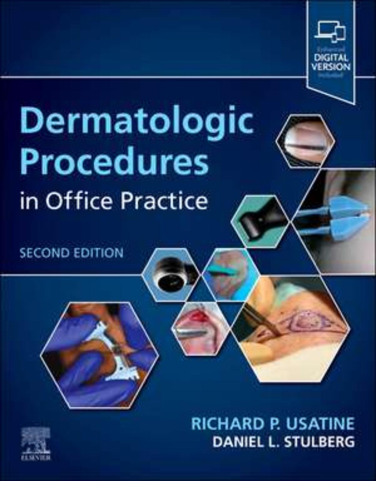
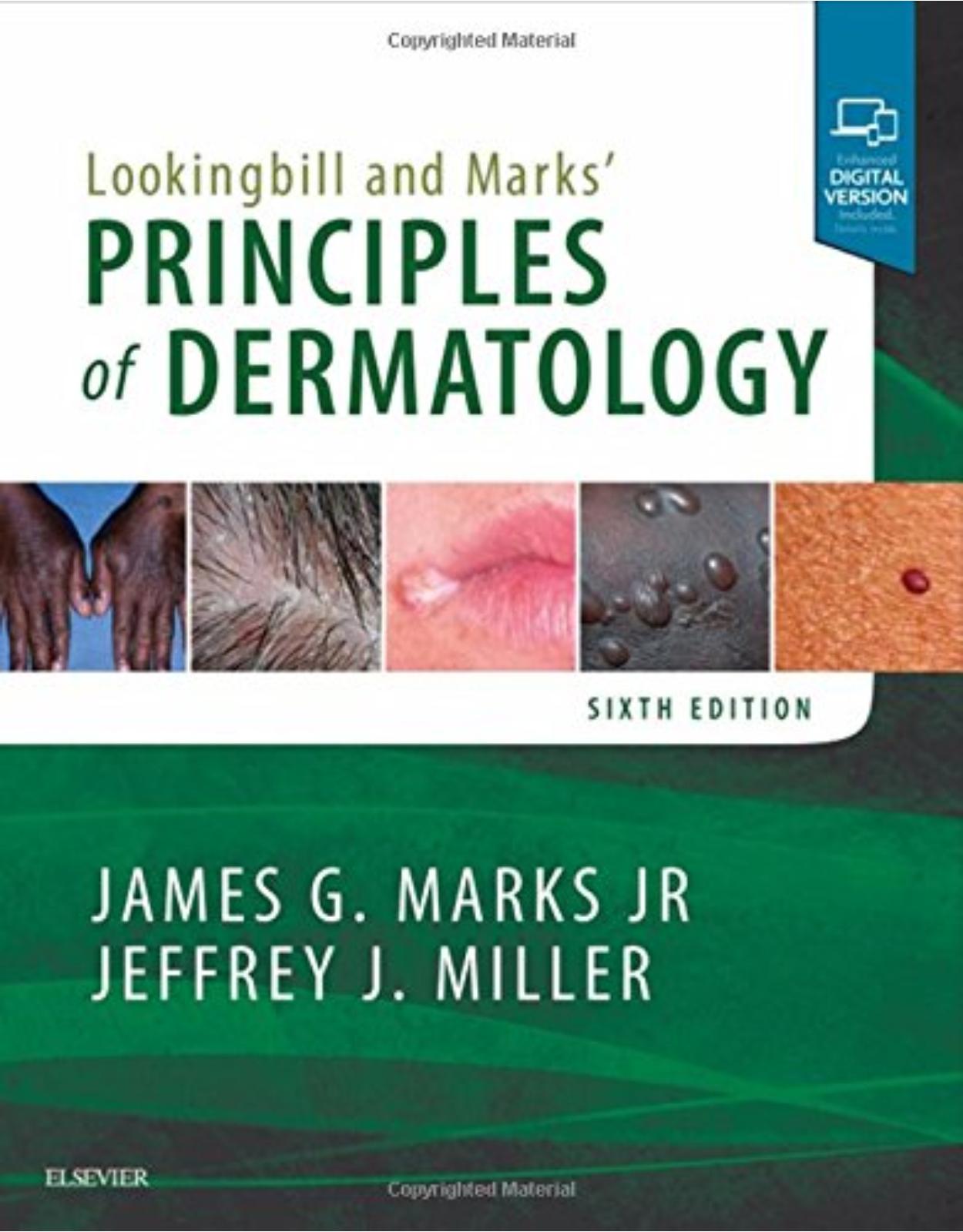
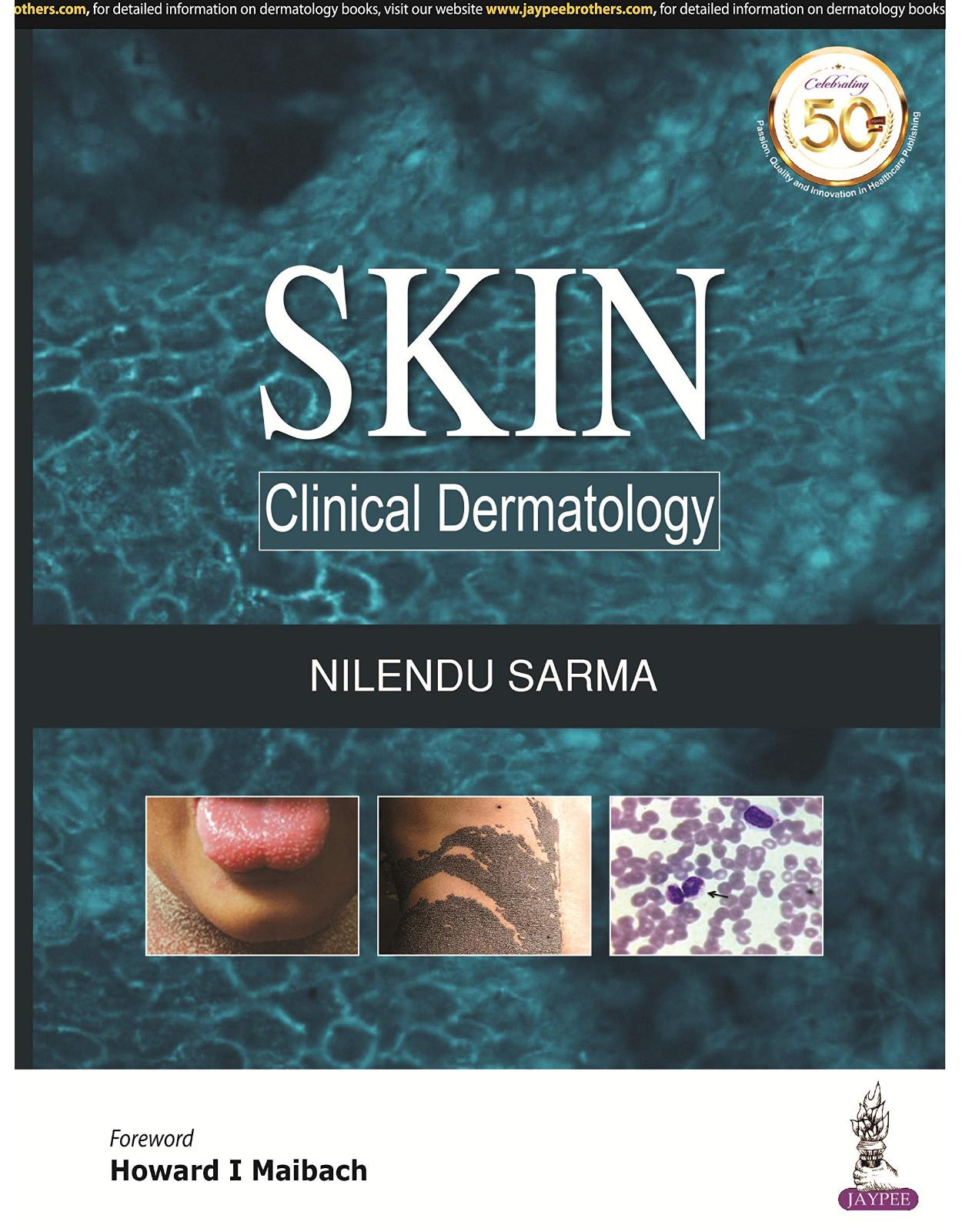
Dermatologic Procedures in Office Practice
Livrare gratis la comenzi peste 500 RON. Pentru celelalte comenzi livrarea este 20 RON.
Disponibilitate: Nu este inca publicata (3-4 saptamani de la data aparitiei)
Editura: Elsevier
Limba: Engleza
Nr. pagini: 560
Coperta: Paperback
Dimensiuni: 216 x 276 mm
An aparitie: iulie 2024
|
Description: |
|
|
|
Covers in-office dermatologic procedures focused on today’s primary care practice. Uses a concise, step-by-step, bulleted format, including set-up, complication avoidance, pearls and pitfalls, and recovery for each procedure. Features an exclusive online library of more than 100 videos, as well as procedural images and patient photographs, photographs of devices and instruments, diagrams, and medical illustrations throughout the text from expert clinicians. Contains new chapters on Biopsies and Excisions in Challenging Locations (face, ears, nose, genital areas) and Dermoscopy of Other Lesions in General Dermatology. Includes handy appendices with sample consent forms, patient education handouts, and a table recommending when to use various procedures based on the diagnosis. An digital version is included with purchase. The digital allows you to access all the text, photographs, figures, and references with the ability to search, customize your content, make notes and highlights, and have content read aloud. |
Table of Contents:
SECTION ONE. Getting Started
1. Preoperative preparation
Surgical planning
Scheduling of complex surgeries
Preoperative medical evaluation
Medical contraindications
Informed consent
Universal precautions
Medications
Standby medications and equipment
Preoperative patient preparation
Drapes
Sterile technique
Time-out
Conclusion
References
2. Setting up your office: Facilities, Instruments, and Equipment
Lighting
Surgical table, stools, and mayo stands
Hand instruments
Hemostasis
Magnification devices
Personal protective equipment
Injectable medications and chemicals
Equipment
What to have in each exam room
Conclusion
Resources
References
3. Anesthesia
Topical anesthetics (Figure 3.1)
EMLA for mucosa (Figure 3.2)
Topical refrigerants/cryoanesthesia
Injection technique – see Videos 3.1–3.3
Dental syringes
Adverse reactions
Debunking the myth of epinephrine-induced necrosis
Nerve blocks – see Video 3.4
Field or ring block – see Video 3.5 and 3.6
Pregnancy and lactation
Conclusion
Summary of basic materials needed
References
4. Hemostasis
Epinephrine – step one
Types of hemostasis
Chemical hemostatic agents
General principles of chemical hemostasis
Electrocoagulation
Electrosurgical equipment and its use
Working in a bloody field
Sterile vs nonsterile procedures
When to use electrocoagulation:
Hemostasis around the eye
Mechanical hemostasis: Pressure and sutures
Buried
Superficial
Oozing on the edges
Using tourniquets, clamps, and instruments to prevent bleeding
Steps to take with nonstop bleeding in surgery
Conclusion
Resources
References
5. Selecting the right suture
Introduction
Wound healing and sutures
Conclusion
Neck and below
If under more tension (back, shoulders, legs)
Resources
References
6. Suturing techniques
Basic skills
Specific suturing techniques
Tips for working with fragile skin
Suture removal
Alternative closure techniques
Learning the techniques: Suggestions on how to practice
Conclusion
References
SECTION TWO. Basic Procedures
7. Laceration repair
Indications for repair
Contraindications for repair
Supplies and equipment
Preprocedure patient preparation
Closure technique
Complications
Postprocedure patient education
Considerations for specific anatomic repairs
CPT/billing codes and ICD-9-CM diagnostic codes
Conclusion
Resources
References
8. Choosing the biopsy type
General principles
Choice of site to biopsy
Is it cancer?
Considerations for specific anatomic areas
How to do a scalp biopsy
How to submit a specimen to the lab
Margin assessment
Conclusion
References
9. The shave biopsy
Indications
Contraindications
Advantages of shave biopsy
Disadvantages of shave biopsy
Equipment (Figure 9.9)
Shave biopsy: Steps and principles
Cutting the shave biopsy
Aftercare
Pathology and follow-up
Suggestion for learning the shave biopsy technique
Less than optimal outcomes
Cosmetic results
Making a diagnosis
Additional examples for shave biopsy
Coding and billing pearls
Conclusion
References
10. The punch biopsy
Indications
Relative contraindications and cautions
Advantages of punch biopsy
Disadvantages of punch biopsy
Equipment
Punch biopsy: Steps and principles
Punch biopsy of a blue nevus (Figure 10.24; see Video 10.6)
Suggestion for learning punch biopsy technique
Complications
Specific examples
Complications
Coding and billing pearls
Conclusion
References
11. The elliptical excision
Indications
Contraindications
Equipment
The elliptical incision: Steps and principles
Notes on infection
Variations
Alternatives to an elliptical excision
Suggestions for learning the elliptical excision technique
Coding and billing pearls
Conclusion
References
12. Cysts and lipomas
Epidermal inclusion cysts (EIC) and variants
Surgical considerations for epidermal inclusion cysts (EICs) and pilar cysts (see Videos 12.1 to 12.5)
General considerations for epidermal cyst or pilar cyst removal
Advantages and disadvantages of various approaches
The procedure
Steps and principles for the minimal excision technique (see Video 12-4)
Pathology
Major alternatives
General considerations in treating digital mucous cysts
Needling and cryotherapy of digital mucous cysts
Alternatives
Complications of any cyst removal technique
Lipomas (see Videos 12.7 to 12.10)
Punch (enucleation) technique to remove lipomas
Coding and billing pearls
Conclusion
References
13. Cryosurgery
Indications
Contraindications
Advantages of cryosurgery
Disadvantages of cryosurgery
Equipment for liquid nitrogen
Cryosurgery: Principles and getting started
Cryosurgery methods
Complications
Treating specific lesions
Learning the techniques see Videos 13.10–13.15
Aftercare
Coding and billing pearls
Conclusion
References
14. Electrosurgery
Advantages of electrosurgery
Disadvantages of electrosurgery
Electrosurgery versus cryosurgery
Electrosurgery versus scalpel
Equipment
Accessories
Indications for use of electrosurgery
Precautions
Electrosurgical techniques: General principles
Safety measures with electrosurgery
Treating specific lesions
Electrodesiccation and curettage
Learning the techniques
Coding and billing pearls
Conclusion
References
15. Intralesional injections
Indications
Contraindications
Equipment
Informed consent
Steroid strength
Treating specific lesions
Complications of steroid injections
Intralesional treatments for warts
Intralesional injections for warts due to HPV
Aftercare for all intralesional injections
Coding and billing pearls
References
16. Incision and drainage
Indications for procedure
Contraindications and cautions
Advantages of incision and drainage
Disadvantages of incision and drainage
Equipment
Procedure: Steps and principles
Aftercare and packing of abscesses
Loop drainage technique
Paronychia
Felon (abscess of the pulp of the distal digit)
Complications of an abscess
Unroofing/deroofing hidradenitis suppurativa
Notes on specific lesions
Special populations
Coding and billing pearls
Conclusion
References
17. Nail procedures
Overview
Anatomy of the nail unit
Nail procedures
Partial nail plate excision
Destruction of nail matrix
Full nail plate removal (nail avulsion)
Biopsies to diagnose pigmented nail changes (acral melanoma versus benign longitudinal melanonychia)
Notes on other lesions
Complications of nail surgery
Algorithm
Nail plate dermatopathology
Postop directions for patients
Coding and billing pearls
Conclusion
References
18. Dermoscopy of skin cancer and benign neoplasia
Advantages of and evidence for dermoscopy
Equipment
Algorithms to learn and perform dermoscopy
TADA
Comedo-like openings (Figure 18.7b)
Fissures and ridges (Figure 18.8)
Two-step dermoscopy algorithm for skin cancer criteria
PRP in persons of color
Conclusion
Resources
References
19. Dermoscopy in general dermatology
Introduction
Inflammatory diseases
Infectious skin diseases
Conclusion
References
SECTION THREE. Advanced Procedures
20. Biopsies and excisions in challenging locations
Scalp
Periocular
Nose
Ears
Lips
Oral
Genitalia
Sole of the foot (see Videos 20.6 and 20.7)
Conclusion
References
21. Flaps
Overview
Terminology
Use of flaps following treatment of malignant tumors
Contraindications
Planning the flap
Advancement flap
Advancement flap
Island pedicle flap
Island pedicle flap
Rotation flap
Single rotation flap
Bilateral O-Z rotation flap
Rotation flap
Transposition flap
Rhombic transposition flap
Zitelli bilobed transposition flap
Transposition flap
Learning the techniques
Coding and billing pearls
References
22. Ultrasound in dermatology
Normal anatomy
Applications
Conclusion
Resources
References
SECTION FOUR. Putting It All Together
23. Procedures to treat benign conditions
Acne surgery
Acrochordons
Angiomas/angiokeratomas/angiofibromas
Angiokeratomas
Angiofibromas
Chondrodermatitis nodularis helicis
Cutaneous horn
Dermatofibromas
Keloids and hypertrophic scars
Lentigines (solar)
Milia
Molluscum contagiosum
Mucocele
Dysplastic nevi (atypical moles)
Shave
Punch excision
Elliptical excision
Dysplastic nevus (atypical mole)
Congenital nevi
Neurofibromas
Pilomatricoma
Pyogenic granuloma (lobular capillary hemangioma)
Sebaceous hyperplasia
Seborrheic keratoses and dermatosis papulosa nigra
Syringomas
Telangiectasias and spider angiomas
Trichoepithelioma
Warts, common
In-office topicals
Curettage and desiccation
Immunotherapy
Special considerations/billing
Warts, filiform
Warts, flat
Warts, plantar
Warts, venereal (condyloma acuminata)
Xanthelasma
Less common benign adnexal tumors
Conclusion
References
24. Diagnosis and treatment of malignant and premalignant lesions
Mohs surgery appropriate use criteria (AUC) app (see Figure 27.13)
Actinic keratoses
Squamous cell carcinoma
Keratoacanthoma
Basal cell carcinoma
Melanoma
Clinical appearance
Cutaneous T-cell lymphomas (including mycosis fungoides)
Dermatofibrosarcoma protuberans
Merkel cell carcinoma
Coding and billing pearls
Conclusion
Resources
Selected guidelines
References
25. Wound care
Wound healing phases
Types of wounds
Pressure injury
Wound care dressings
Wound care
Human amniotic membranes
Prevention of pressure injuries
Treatment of pressure injuries
Special circumstances
Postoperative wound care patient instructions
Special locations
Complications
Conclusion
References
26. Complications: Postprocedural Adverse Effects and Their Prevention
Calling patients to check on them after a large procedure
Responding quickly to patients’ concerns after a procedure
Review of the literature
Clinical management and evaluation of possible infection
Pain
Bleeding
Swelling and bruising
Scarring
Wound dehiscence
Pigmentation changes
Flap necrosis
Nerve damage
Recurrence of a lesion or skin cancer
Making mistakes
Conclusion
References
27. When to refer/Mohs surgery
Mohs surgery
Appropriate use criteria guideline and a free app
Radiation
Systemic agents for severe forms of BCC and SCC
Referral and systemic therapies for advanced melanoma
Conclusion
References
28. Coding common skin procedures
Comparing medical work
Biopsies
Destructions of lesions (see Chapters 13, 14)
Shave removal
Excisions (see Chapter 11, elliptical excision, and Chapter 12, cysts and lipomas)
Skin repairs (see Chapter 7, laceration repair, and Chapter 11, elliptical excision)
Incision and drainage (see Chapter 16) (Table 28.11)
Foreign body removal (Table 28.12)
Cosmetic removals
Measure, review, and learn
References
Appendix A. Consent forms and patient information handouts
Appendix B. Procedures to consider for benign, premalignant, and malignant conditions
Index
| An aparitie | iulie 2024 |
| Autor | Richard P. Usatine, Daniel L. Stulberg |
| Dimensiuni | 216 x 276 mm |
| Editura | Elsevier |
| Format | Paperback |
| ISBN | 9780323930628 |
| Limba | Engleza |
| Nr pag | 560 |
| Versiune digitala | DA |
-
 1,21200 lei 1,10000 lei
Clientii ebookshop.ro nu au adaugat inca opinii pentru acest produs. Fii primul care adauga o parere, folosind formularul de mai jos.