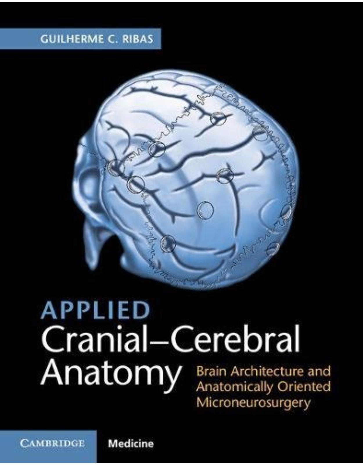
Applied Cranial-Cerebral Anatomy
Livrare gratis la comenzi peste 500 RON. Pentru celelalte comenzi livrarea este 20 RON.
Disponibilitate: La comanda in aproximativ 4-6 saptamani
Autor: Guilherme C. Ribas
Editura: Cambridge
Limba: Engleza
Nr. pagini: 140
Coperta: Hardback
Dimensiuni: 21.9 x 1.2 x 27.6 cm
An aparitie: 2018
Description:
This book is the first to offer a comprehensive guide to understanding the brain's architecture from a topographical viewpoint. Authored by a leading expert in surgical neuroanatomy, this practical text provides tri-dimensional understanding of the cerebral hemispheres, and the relationships between cerebral surfaces and the skull's outer surfaces through detailed brain dissections and actual clinical cases with operative photographs and correlative neuroimaging. For neurosurgeons, neuroradiologists and neurologists at all levels, this book emphasises the anatomy of the sulci and gyri of the cerebral surface. It is an essential resource for the general neurosurgery practice, and more particularly for planning surgical access routes for intracranial tumors.
Table of Contents:
Chapter 1 Historical Remarks
1.1 The Cerebral Surface
1.2 Cerebral Cortical Cytoarchitecture
1.3 White Matter Fibers
1.4 Cranial-Cerebral Relationships
1.5 Technology and Cerebral Localization
1.6 Microneurosurgical Anatomy
Chapter 2 The Cerebral Architecture
2.1 Developmental Aspects
2.1.1 Evolutionary Considerations
2.1.2 Embryological and Fetal Considerations
2.2 The Cerebral Hemispheres
2.2.1 The Meninges, the Subarachnoid Space, and the Main Cerebral Fissures
2.2.2 The Cerebral Surface, Its Sulci and Gyri
2.2.3 The Cerebral Lobes and Related Regions
2.2.3.1 The Frontal Lobe
2.2.3.1.1 Frontal Lobe Sulci and Gyri
2.2.3.2 Parietal Lobe
2.2.3.2.1 Parietal Lobe Sulci and Gyri
2.2.3.3 The Occipital Lobe
2.2.3.3.1 Occipital Lobe Sulci and Gyri
2.2.3.4 The Temporal Lobe
2.2.3.4.1 The Temporal Lobe Sulci and Gyri
2.2.3.5 The Insular Lobe and the Central Core
2.2.3.5.1 Insular Lobe Sulci and Gyri
2.2.3.5.2 The Insula and the Cerebral Central Core
2.2.3.6 The Limbic Lobe and Correlated Areas
2.2.3.6.1 Limbic Lobe Sulci and Gyri
2.2.3.6.2 The Temporal Stem and the Sagittal Stratum
2.2.3.6.3 The Basal Forebrain: the Anterior Perforated Substance, the Ventral-Striato-Pallidal and Septal Regions
2.2.3.6.4 The Limbic System
2.2.4 The White Matter of the Cerebral Hemispheres
2.2.4.1 Association Fibers
2.2.4.2 Superior Longitudinal Fasciculus
2.2.4.3 Inferior Fronto-Occipital Fasciculus
2.2.4.4 Uncinate Fasciculus
2.2.4.5 Inferior Longitudinal Fasciculus
2.2.4.6 Cingulum
2.2.5 Commissural Fibers
2.2.5.1 Corpus Callosum
2.2.5.2 Anterior Commissure
2.2.5.3 Hippocampal Commissure
2.2.5.4 Posterior Commissure
2.2.5.5 Habenular Commissure
2.2.6 Projection Fibers
2.2.6.1 Internal Capsule
Chapter 3 Cranial-Cerebral Relationships Applied to Microneurosurgery
3.1 Microneurosurgical Anatomy – General Remarks
3.2 The Sulcal, Gyral, and Cranial Key Points
3.2.1 The Concept of Sulcal and Gyral Key Points and Their Cranial-Cerebral Relationships
3.2.2 Fronto-Opercular Key Points
3.2.2.1 The Anterior Sylvian Point
3.2.2.2 The Inferior Rolandic Point
3.2.2.3 The Inferior Frontal and Precentral Sulci Meeting Point
3.2.2.4 The Frontoparietal Operculum
3.2.2.5 Frontotemporal Craniotomies and Exposures
3.2.2.6 Anatomical Remarks Pertinent to Common Frontotemporal Transcerebral Procedures
Approaches to the Temporal Horn of the Lateral Ventricle
Anterior Temporal Lobectomy and Lateral Approaches for Exposure of the Temporal Horn
Transsylvian Approaches to the Temporal Horn
Basal Approaches
3.2.3 Superior Frontal and Central Key Points
3.2.3.1 The Superior Frontal and Precentral Sulci Meeting Point
3.2.3.2 The Superior Rolandic Point
3.2.4 Superior Frontal and Central Craniotomies
3.2.4.1 Anatomical Remarks Pertinent to Common Frontal Transcerebral Procedures
Exposure of the Superior Frontal Gyrus and Sulcus, and of the Interhemispheric Fissure
Transcallosal Approaches to the Lateral Ventricles
Transcallosal Approaches to the Third Ventricle
3.2.5 Parietal Key Points
3.2.5.1 The Intraparietal and Postcentral Sulci Meeting Point
3.2.5.2 The Euryon and the Supramarginal Gyrus
3.2.5.3 The Parieto-Occipital Incisure and the Lambda
3.2.5.4 Parietal Craniotomies
3.2.5.5 Anatomical Remarks Pertinent to Common Parietal Transcerebral Procedures
Transparietal Approach to the Atrium
3.2.6 Posterior Temporal Key Point
3.2.6.1 The Posterior Extremity of the Superior Temporal Sulcus
3.2.6.2 Posterior Temporal Craniotomies
3.2.6.3 Anatomical Remarks Pertinent to Posterior Temporal Approaches
3.2.7 Occipital Key Point
3.2.7.1 The Opisthocranion
3.2.7.2 Occipital Craniotomies
3.2.7.3 Anatomical Remarks Pertinent to Common Occipital Transcerebral Procedures
Occipital Lobectomy
Occipital Interhemispheric Approach to the Atrium
3.3 The Basal Supratentorial Key Points
3.3.1 The Frontozygomatic Process Key Point
3.3.2 The Anterior Temporal Key Point
3.3.3 The Preauricular Depression Key Point
3.3.4 The Parietomastoid and Squamosal Suture Meeting Point
3.3.5 The Asterion
References
Index
| An aparitie | 2018 |
| Autor | Guilherme C. Ribas |
| Dimensiuni | 21.9 x 1.2 x 27.6 cm |
| Editura | Cambridge |
| Format | Hardback |
| ISBN | 9781107156784 |
| Limba | Engleza |
| Nr pag | 140 |












Clientii ebookshop.ro nu au adaugat inca opinii pentru acest produs. Fii primul care adauga o parere, folosind formularul de mai jos.