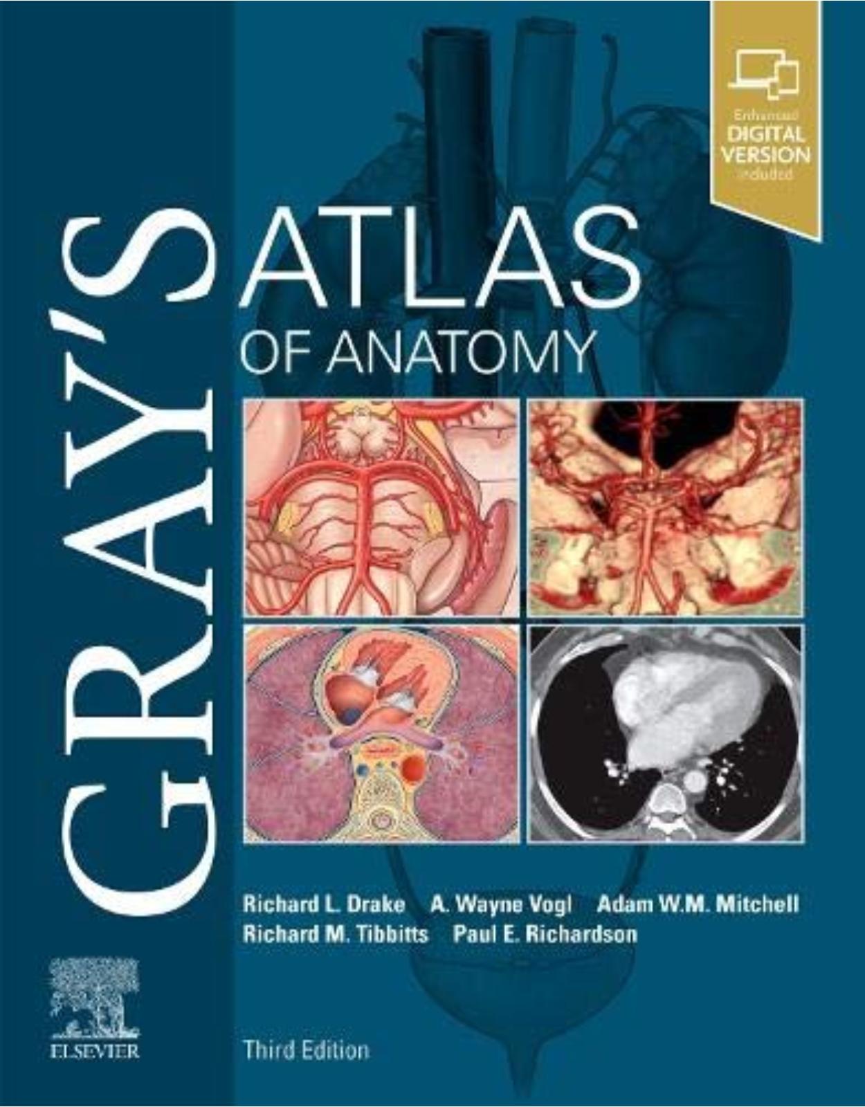
Gray’s Atlas of Anatomy, 3rd Edition
Livrare gratis la comenzi peste 500 RON. Pentru celelalte comenzi livrarea este 20 RON.
Disponibilitate: La comanda in aproximativ 4 saptamani
Editura: Elsevier
Limba: Engleza
Nr. pagini: 648
Coperta: Paperback
Dimensiuni: 21.59 x 2.79 x 27.43 cm
An aparitie: 9 April 2020
Description:
Clinically focused, consistently and clearly illustrated, and logically organized, Gray's Atlas of Anatomy, the companion resource to the popular Gray's Anatomy for Students, presents a vivid, visual depiction of anatomical structures. Stunning illustrations demonstrate the correlation of structures with clinical images and surface anatomy - essential for proper identification in the dissection lab and successful preparation for course exams.
Build on your existing anatomy knowledge with structures presented from a superficial to deep orientation, representing a logical progression through the body.
Identify the various anatomical structures of the body and better understand their relationships to each other with the visual guidance of nearly 1,000 exquisitely illustrated anatomical figures.
Visualize the clinical correlation between anatomical structures and surface landmarks with surface anatomy photographs overlaid with anatomical drawings.
Recognize anatomical structures as they present in practice through more than 270 clinical images - including laparoscopic, radiologic, surgical, ophthalmoscopic, otoscopic, and other clinical views - placed adjacent to anatomic artwork for side-by-side comparison.
Gain a more complete understanding of the inguinal region in women through a brand-new, large-format illustration, as well as new imaging figures that reflect anatomy as viewed in the modern clinical setting.
Table of Contents:
Chapter 1: THE BODY
CONTENTS
Anatomical position, terms, and planes
Anatomical planes and imaging
Surface anatomy: anterior view
Surface anatomy: posterior view
Skeleton: anterior
Skeleton: posterior
Muscles: anterior
Muscles: posterior
Vascular system: arteries
Vascular system: veins
Lymphatic system
Nervous system
Sympathetics
Parasympathetics
Dermatomes
Cutaneous nerves
Chapter 2: BACK
CONTENTS
Surface anatomy
Vertebral column
Regional vertebrae
Cervical vertebrae
Thoracic vertebrae
Lumbar vertebrae
Sacrum
Intervertebral foramina and discs
Intervertebral disc problems
Joints and ligaments
Back musculature: surface anatomy
Superficial musculature
Intermediate musculature
Deep musculature
Back musculature: transverse section
Suboccipital region
Spinal nerves
Spinal cord
Spinal cord vasculature
Venous drainage of spinal cord
Meninges
Spinal cord: imaging
Transverse section: thoracic region
Dermatomes and cutaneous nerves
Tables
Chapter 3: THORAX
CONTENTS
Surface anatomy with bones
Bony framework
Ribs
Articulations
Breast
Pectoral region
Thoracic wall muscles
Diaphragm
Arteries of the thoracic wall
Veins of the thoracic wall
Nerves of the thoracic wall
Lymphatics of the thoracic wall
Intercostal nerves and arteries
Pleural cavities and mediastinum
Parietal pleura
Surface projections of pleural recesses
Right lung
Left lung
Lung lobes: surface relationship
Lung lobes: imaging
Bronchial tree
Bronchopulmonary segments
Pulmonary vessels and plexus
Pulmonary vessels: imaging
Mediastinum
Pericardium
Pericardial layers
Anterior surface of heart
Base and diaphragmatic surface of heart
Right atrium
Right ventricle
Left atrium
Left ventricle
Aortic valve and cardiac skeleton
Cardiac chambers and heart valves
Coronary vessels
Coronary arteries and variations
Cardiac conduction system
Auscultation points and heart sounds
Cardiac innervation
Superior mediastinum: thymus
Superior mediastinum: veins and arteries
Superior mediastinum: arteries and nerves
Superior mediastinum: imaging
Superior mediastinum: veins and trachea
Mediastinum: imaging
Mediastinum: view from right
Mediastinum: imaging – view from right
Mediastinum: view from left
Mediastinum: imaging – view from left
Posterior mediastinum
Mediastinum: imaging
Transverse section: TVIII level
Dermatomes and cutaneous nerves
Visceral efferent (motor) innervation of the heart
Visceral afferents
Tables
Chapter 4: ABDOMEN
CONTENTS
Surface anatomy
Quadrants and regions
Abdominal wall
Muscles
Muscles: rectus sheath
Vessels of the abdominal wall
Arteries and lymphatics of the abdominal wall
Nerves of the abdominal wall
Dermatomes and cutaneous nerves
Inguinal region
Inguinal canal in men
Inguinal canal in women
Inguinal hernias
Anterior abdominal wall
Greater omentum
Abdominal viscera
Peritoneal cavity
Abdominal sagittal section
Abdominal coronal section
Arterial supply of viscera
Stomach
Spleen
Arteries of stomach and spleen
Duodenum
Small intestine
Large intestine
Ileocecal junction
Gastrointestinal tract: imaging
Mesenteric arteries
Liver
Vessels of the liver
Segments of the liver
Pancreas and gallbladder
Vasculature of pancreas and duodenum
Venous drainage of viscera
Portosystemic anastomoses
Posterior wall
Vessels of the posterior wall
Diaphragm
Kidneys
Gross structure of kidneys
Kidneys: imaging
Renal vasculature
Branches of the abdominal aorta
Inferior vena cava
Abdominal aorta and inferior vena cava: imaging
Lumbar plexus
Lumbar plexus: cutaneous distribution
Lymphatics
Abdominal innervation
Splanchnic nerves
Visceral efferent (motor) innervation diagram
Visceral afferent (sensory) innervation and referred pain diagram
Kidney and ureter visceral afferent (sensory) diagram
Tables
Chapter 5: PELVIS AND PERINEUM
Surface anatomy and articulated pelvis in men
Surface anatomy and articulated pelvis in women
Pelvic girdle
Pelvic girdle: imaging
Lumbosacral joint
Sacro-iliac joint
Pelvic inlet and outlet
Orientation of pelvic girdle and pelvic brim
Pelvic viscera and perineum in men
Pelvic viscera and perineum in men: imaging
Pelvic viscera and perineum in women
Pelvic viscera and perineum in women: imaging
Lateral wall of pelvic cavity
Floor of pelvic cavity: pelvic diaphragm
Rectum and bladder in situ
Rectum
Bladder in men
Bladder in women
Reproductive system in men
Prostate
Prostate and seminal vesicles
Scrotum
Testes
Penis
Reproductive system in women
Uterus and ovaries
Uterus
Uterus: imaging
Pelvic fascia
Arterial supply of pelvis
Venous drainage of pelvis
Vasculature of the pelvic viscera
Vasculature of uterus
Venous drainage of prostate and penis
Venous drainage of rectum
Sacral and coccygeal nerve plexuses
Pelvic nerve plexus
Hypogastric plexus
Surface anatomy of the perineum
Borders and ceiling of the perineum
Deep pouch and perineal membrane
Muscles and erectile tissues in men
Erectile tissue in men: imaging
Muscles and erectile tissues in women
Erectile tissue in women: imaging
Internal pudendal artery and vein
Pudendal nerve
Vasculature of perineum
Nerves of perineum
Lymphatics of pelvis and perineum in men
Lymphatics of pelvis and perineum in women
Lymphatics
Dermatomes
Innervation of reproductive system in men
Innervation of reproductive system in women
Innervation of bladder
Pelvic cavity imaging in men
Pelvic cavity imaging in women
Tables
Chapter 6: LOWER LIMB
CONTENTS
Surface anatomy
Bones of the lower limb
Pelvic bones and sacrum
Articulated pelvis
Proximal femur
Hip joint
Hip joint: structure and arterial supply
Gluteal region: attachments and superficial musculature
Gluteal region: superficial and deep muscles
Gluteal region: arteries and nerves
Distal femur and proximal tibia and fibula
Thigh: muscle attachments
Thigh: anterior superficial musculature
Thigh: posterior superficial musculature
Thigh: anterior compartment muscles
Thigh: medial compartment muscles
Femoral triangle
Anterior thigh: arteries and nerves
Anterior thigh: arteries
Thigh: posterior compartment muscles
Posterior thigh: arteries and nerves
Transverse sections: thigh
Knee joint
Ligaments of the knee
Menisci and cruciate ligaments
Knee: bursa and capsule
Knee surface: muscles, capsule, and arteries
Popliteal fossa
Tibia and fibula
Bones of the foot
Bones and joints of the foot
Talus and calcaneus
Ankle joint
Ligaments of the ankle joint
Leg: muscle attachments
Posterior leg: superficial muscles
Posterior compartment: deep muscles
Posterior leg: arteries and nerves
Lateral compartment: muscles
Anterior leg: superficial muscles
Anterior compartment: muscles
Anterior leg: arteries and nerves
Leg: cutaneous nerves
Transverse sections: leg
Foot: muscle attachments
Foot: ligaments
Dorsum of foot
Dorsum of foot: arteries and nerves
Plantar aponeurosis
Plantar region (sole) musculature: first layer
Plantar region (sole) musculature: second layer
Plantar region (sole) musculature: third layer
Plantar region (sole) musculature: fourth layer
Plantar region (sole): arteries and nerves
Dorsal hood and tarsal tunnel
Superficial veins of the lower limb
Lymphatics of the lower limb
Anterior cutaneous nerves and dermatomes of the lower limb
Posterior cutaneous nerves and dermatomes of the lower limb
Tables
Chapter 7: UPPER LIMB
CONTENTS
Surface anatomy
Bones of the upper limb
Bony framework of shoulder
Scapula
Clavicle: joints and ligaments
Proximal humerus
Glenohumeral joint
Muscle attachments
Pectoral region
Deep pectoral region
Walls of the axilla
The four rotator cuff muscles
Deep vessels and nerves of the shoulder
Axillary artery
Brachial artery
Brachial plexus
Medial and lateral cords
Posterior cord
Distal end of humerus and proximal end of radius and ulna
Muscle attachments
Anterior compartment: muscles
Anterior compartment: arteries and nerves
Veins of the arm
Posterior compartment: muscles
Posterior compartment: arteries and nerves
Lymphatics of the arm
Transverse sections: arm
Anterior cutaneous nerves of the arm
Posterior cutaneous nerves of the arm
Elbow joint
Elbow joint: capsule and ligaments
Cubital fossa
Radius and ulna
Bones of the hand and wrist joint
Imaging of the hand and wrist joint
Bones of the hand
Joints and ligaments of the hand
Muscle attachments of forearm
Anterior compartment of forearm: muscles
Anterior compartment of forearm: arteries and nerves
Posterior compartment of forearm: muscles
Posterior compartment of forearm: arteries and nerves
Transverse sections: forearm
Carpal tunnel
Muscle attachments of the hand
Superficial palmar region (palm) of hand
Tendon sheaths of hand
Lumbrical muscles
Intrinsic muscles of hand
Palmar region (palm) of hand: arteries and nerves
Arteries of the hand
Innervation of the hand: median and ulnar nerves
Dorsum of hand
Dorsal hoods
Dorsum of hand: arteries
Dorsum of hand: nerves
Anatomical snuffbox
Superficial veins and lymphatics of forearm
Anterior cutaneous nerves of forearm
Posterior cutaneous nerves of upper limb
Tables
Chapter 8: HEAD AND NECK
CONTENTS
Surface anatomy with bones
Bones of the skull
Skull: anterior view
Skull: lateral view
Skull: posterior view
Skull: superior view and roof
Skull: inferior view
Skull: cranial cavity
Ethmoid, lacrimal bone, inferior concha, and vomer
Maxilla and palatine bone
Skull: muscle attachments
Scalp and meninges
Dural partitions
Dural arteries and nerves
Dural venous sinuses
Brain
Brain: imaging
Cranial nerves
Arterial supply to brain
Cutaneous distribution of trigeminal nerve [V]
Facial muscles
Vasculature, facial nerve [VII] and lymphatics
Deep arteries and veins of parotid region
Bony orbit
Section through orbit and structures of eyelid
Eyelids and lacrimal apparatus
Innervation of the lacrimal gland
Muscles of the eyeball
Innervation of the orbit and eyeball
Eye movements
Vasculature of orbit
Eyeball
Eye imaging
Ear surface and sensory innervation
Ear
Middle ear
Internal ear
Ear imaging
Temporal and infratemporal fossae
Bones of the temporal and infratemporal fossae
Temporal and infratemporal fossae
Temporomandibular joint
Mandibular division of the trigeminal nerve [V]
Parasympathetic innervation
Arteries and veins of temporal and infratemporal fossae
Pterygopalatine fossa
Neck surface anatomy
Bones of the neck
Compartments and fascia of the neck
Superficial veins of the neck
Muscles of the neck
Nerves in the neck
Cranial nerves in the neck
Cervical plexus and sympathetic trunk
Arteries of the neck
Root of the neck: arteries
Lymphatics of the neck
Pharynx
Muscles of the pharynx
Innervation of the pharynx
Vasculature of the pharynx
Larynx
Laryngeal cavity
Muscles of the larynx
Innervation of the larynx
Thyroid gland
Vasculature of the thyroid gland
Nose and paranasal sinuses
Nasal cavity: bones
Nasal cavity: mucosal linings
Vasculature and innervation of the nasal cavity
Sinus imaging
Oral cavity: bones
Teeth
Teeth: imaging
Anatomy of teeth
Vessels and nerves supplying teeth
Innervation of teeth and gums
Muscles and salivary glands of the oral cavity
Vessels and nerves of the tongue
Tongue
Hard and soft palate
Palate
Innervation of oral cavity
Cranial nerves
Visceral motor pathways in the head
Tables
INDEX
| An aparitie | 9 April 2020 |
| Autor | Richard Drake PhD FAAA , A. Wayne Vogl PhD FAAA , Adam W. M. Mitchell MB BS FRCS FRCR |
| Dimensiuni | 21.59 x 2.79 x 27.43 cm |
| Editura | Elsevier |
| Format | Paperback |
| ISBN | 9780323636391 |
| Limba | Engleza |
| Nr pag | 648 |
-
30900 lei 26000 lei









Clientii ebookshop.ro nu au adaugat inca opinii pentru acest produs. Fii primul care adauga o parere, folosind formularul de mai jos.