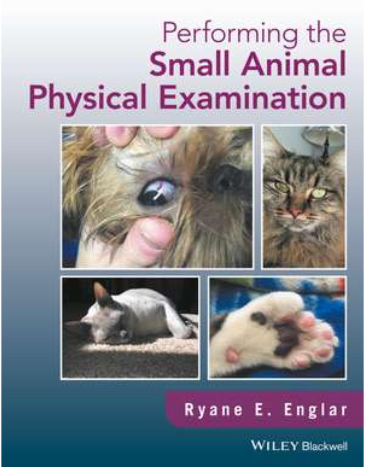
Performing the Small Animal Physical Examination
Livrare gratis la comenzi peste 500 RON. Pentru celelalte comenzi livrarea este 20 RON.
DESCRIPTION
Performing the Small Animal Physical Examination offers an easy-to-follow guide to successfully executing a thorough physical exam in cats and dogs, with nearly 1,000 clinical photographs depicting step-by-step details.
· Provides comprehensive, practical information on the physical examination in small animal patients
· Presents nearly 1,000 color photographs with step-by-step details of the procedures and principles
· Offers advice on preparing the examination room, useful tips, and concrete guidance for examining each body system
· Outlines a systematic, in-depth approach to the initial examination in dogs and cats
· Supports new and experienced veterinarians and veterinary technicians alike in performing a thorough basic exam
TABLE OF CONTENTS
About the Author xiii
Preface xiv
Acknowledgments xv
Part One Performing the Feline Physical Examination 1
1 Setting the Stage: Feline-Friendly Practice 3
1.1 Challenges Faced in Feline Practice 3
1.2 The Emergence of Feline-Friendly Practice 4
1.3 Key Principles of Feline-Friendly Practice 4
1.4 The Role of Sound 8
1.5 The Role of Tactile Stimulation 8
1.6 The Role of Scent 8
1.7 The Role of Advance Preparation 9
1.8 Examination Room Etiquette: Accessing the Cat 10
1.9 Recognizing Body Language 12
1.10 Feline-Friendly Handling 16
1.11 Other Feline Handling Tools 20
2 Assessing the Big Picture: the Body, the Coat, and the Skin of the Cat 24
2.1 Forms of Identification 24
2.2 Body Condition Scoring 25
2.3 Assessing Hydration 29
2.4 Inspecting the Coat: First Impressions 30
2.5 Identifying Coat Colors and Coat Patterns 32
2.6 Assessing Coat Quality 34
2.7 Inspecting the Skin 40
3 Examining the Head of the Cat 52
3.1 Skull Shape and Facial Symmetry 52
3.2 The Eyes and Accessory Visual Structures 52
3.2.1 A Systematic Approach to the Eye Examination 52
3.2.2 Evaluating the Adnexa of the Eye 53
3.2.3 Evaluating the Globe 58
3.2.4 Evaluating the Sclera 60
3.2.5 Evaluating the Cornea 61
3.2.6 Evaluating the Iris 61
3.2.7 Evaluating the Pupils 62
3.2.8 Assessing Ocular Reflexes 62
3.2.9 Assessing the Anterior Chamber 64
3.2.10 Assessing the Lens 65
3.2.11 Introduction to Fundoscopy 66
3.2.12 Fundoscopy and Direct Ophthalmoscopy 67
3.2.13 Fundoscopy and Indirect Ophthalmoscopy 67
3.3 The Ears 68
3.4 The Nose 72
3.5 The Extra-Oral Examination 73
3.6 The Intra-Oral Examination 75
3.6.1 Assessing Mucous Membrane Color 75
3.6.2 Assessing Capillary Refill Time 76
3.6.3 Examining the Mucosa 76
3.6.4 Examining the Gingiva 78
3.6.5 Assessing the Dentition 78
3.6.6 Assessing the Occlusion 79
3.6.7 Assessing for Calculus 81
3.6.8 Opening the Mouth 81
3.6.9 Examining the Tongue 82
3.6.10 Assessing for Periodontal Disease 82
3.6.11 Feline-Specific Dentistry 84
4 Examining the Endocrine and Lymphatic Systems of the Cat 90
4.1 Evaluating the Thyroid Gland 90
4.1.1 The Pathophysiology of Hyperthyroidism 90
4.1.2 The Etiology of Hyperthyroidism 91
4.1.3 The Art of Palpating an Enlarged Thyroid Gland 91
4.2 Assessing the Lymphatic System 93
4.2.1 Examining the Submandibular Lymph Nodes 93
4.2.2 Examining the Superficial Cervical or Pre-Scapular Lymph Nodes 93
4.2.3 Examining the Popliteal Lymph Nodes 94
4.2.4 Feeling for Lymph Nodes That Should Not Be Present 94
5 Examining the Cardiovascular and Respiratory Systems of the Cat 98
5.1 The Cardiac Patient 98
5.2 Assessing the Cardiovascular System Prior to Auscultation 99
5.2.1 Attitude 99
5.2.2 Respiratory Rate 99
5.2.3 Respiratory Effort 99
5.2.4 Respiratory Route 99
5.2.5 Mucous Membrane Color 99
5.2.6 Capillary Refill Time (CRT) 99
5.2.7 Jugular Pulse 99
5.2.8 Palpating the Ventral Neck 100
5.2.9 Palpating the Limbs for Warmth and Assessing the Extremities for Color 100
5.2.10 Assessing Femoral Pulses 100
5.3 Cardiothoracic Auscultation 101
5.3.1 Recalling the Cardiac Cycle 101
5.3.2 Normal Heart Sounds 101
5.3.3 Abnormal Heart Sounds: Murmurs 101
5.3.4 Other Heart Sounds 102
5.3.5 Ausculting the Heart 102
5.3.6 Understanding How the Stethoscope Is Built to Facilitate Auscultation 105
5.4 The Respiratory Patient 105
5.4.1 The Upper Airway Patient 105
5.4.2 The Lower Airway Patient 105
5.4.3 The Patient with Thoracic Cavity Disease 107
5.5 Assessing the Respiratory System Prior to Auscultation 110
5.5.1 The Nose 112
5.5.2 The Larynx and the Trachea 113
5.5.3 Thoracic Compliance 113
5.5.4 Thoracic Percussion 113
5.6 Understanding Normal Airway Sounds 114
5.7 Ausculting the Airway 114
5.8 Understanding Adventitious Airway Sounds 115
5.9 Using Airway Sounds to Corroborate Percussive Findings 116
5.10 Purring as an Obstruction to Auscultation 116
6 Examining the Abdominal Cavity of the Cat 120
6.1 Overview of the Digestive Tract as It Pertains to Presenting Complaints 120
6.2 The Esophagus 122
6.3 Visual Inspection of the Abdomen 122
6.4 Superficial Palpation of the Abdomen 122
6.5 Deep Palpation of the Abdomen 124
6.5.1 The Liver 126
6.5.2 The Stomach 127
6.5.3 The Spleen 127
6.5.4 The Pancreas 127
6.5.5 The Small Intestine 128
6.5.6 Mesenteric Lymph Nodes 129
6.5.7 The Large Intestine 129
6.5.8 The Rectal Examination 130
6.6 The Upper Urinary Tract 132
6.7 The Lower Urinary Tract 134
6.8 The Male Reproductive Tract 135
6.9 The Female Reproductive Tract 137
6.10 Being Presented with a Female of Unknown Sexual Status 138
6.11 Neonates 138
7 Examining the Musculoskeletal System of the Cat 145
7.1 Muscle Condition Score (MCS) 145
7.2 The Skeleton as a Whole 146
7.2.1 Key Components of the Axial Skeleton to Appreciate on Physical Examination 148
7.2.2 Key Components of the Appendicular Skeleton to Appreciate on Physical Examination 150
7.2.3 Additional Components of the Skeleton to Appreciate on Physical Examination 153
7.3 The Appendicular Skeleton: The Forelimb 153
7.4 The Appendicular Skeleton: The Hind Limb 160
8 Evaluating the Nervous System of the Cat 174
8.1 Assessing Behavior and Mental Status 174
8.2 Assessing Posture 176
8.3 Assessing Coordination and Gait 177
8.4 Assessing Postural Reactions 178
8.5 Assessing for Other Abnormal Movements 180
8.6 Evaluating the Spinal Reflexes 181
8.7 Assessing the Cranial Nerves 183
8.7.1 Reviewing the Ocular Reflexes Associated with the Cranial Nerves 183
8.7.2 Reviewing the Cranial Nerves Associated with Ocular Movement 185
8.7.3 Reviewing the Cranial Nerves Associated with Tactile Sensation 185
8.7.4 Reviewing the Cranial Nerves Associated with Muscle Movement Other Than Ocular 186
8.7.5 Reviewing the Cranial Nerves Associated with Digestion 186
8.7.6 Reviewing the Cranial Nerves Associated with Maintaining Posture 186
8.8 Assessing Nociception 186
Part Two Performing the Canine Physical Examination 191
9 Setting the Stage: Canine-Friendly Practice and Low-Stress Handling 193
9.1 Challenges Faced in Canine Practice 193
9.2 The Concept of Low-Stress Handling 194
9.3 White Coat Syndrome 196
9.4 The Role of Scent 198
9.5 The Role of Advance Preparation 199
9.6 Examination Room Etiquette: Setting the Tone for Initial Veterinary Interactions with the Dog 199
9.7 Recognizing Body Language 199
9.8 Creative Approaches to Challenging Interactions with Canine Patients 205
9.9 Other Canine Handling Tools 206
10 Assessing the Big Picture: the Body, the Coat, and the Skin of the Dog 213
10.1 Forms of Identification 213
10.2 Body Condition Scoring 214
10.3 Assessing Hydration 221
10.4 Breed Designation 222
10.5 Inspecting the Coat: First Impressions 223
10.6 Identifying Coat Colors and Coat Patterns 225
10.7 Assessing Coat Quality 233
10.8 Inspecting the Skin 237
10.9 Primary Skin Lesions 240
10.10 Secondary Skin Lesions 245
10.11 Miscellaneous Skin Lesions 246
10.12 Hyperkeratosis 249
10.13 Skin Folds 250
10.14 Nails and Paw Pads 251
10.15 Skin Incisions 252
10.16 Mammary Glands 255
11 Examining the Head of the Dog 261
11.1 Skull Shape: Function Versus Cosmesis 261
11.2 Facial symmetry 265
11.3 The Eyes and Accessory Visual Structures 265
11.3.1 A Systematic Approach to the Eye Examination 265
11.3.2 Evaluating the Adnexa of the Eye 265
11.3.3 Evaluating the Globe 274
11.3.4 Evaluating the Sclera 275
11.3.5 Evaluating the Cornea 277
11.3.6 Evaluating the Iris 278
11.3.7 Evaluating the Pupils 280
11.3.8 Assessing Ocular Reflexes 281
11.3.9 Assessing the Anterior Chamber 281
11.3.10 Assessing the Lens 282
11.3.11 Introduction to Fundoscopy 282
11.3.12 Fundoscopy and Direct Ophthalmoscopy 283
11.3.13 Fundoscopy and Indirect Ophthalmoscopy 283
11.4 The Ears 284
11.5 The Nose 289
11.6 The Extra-Oral Examination 291
11.7 The Intra-Oral Examination 295
11.7.1 Assessing Mucous Membrane Color 295
11.7.2 Assessing Capillary Refill Time 296
11.7.3 Examining the Mucosa 296
11.7.4 Examining the Gingiva 296
11.7.5 Assessing the Dentition 297
11.7.6 Assessing the Occlusion 301
11.7.7 Assessing for Calculus 302
11.7.8 Miscellaneous Acquired Tooth-Related Defects 303
11.7.9 Opening the Mouth 304
11.7.10 Examining the Tongue 305
11.7.11 Assessing for Periodontal Disease 306
12 Examining the Endocrine and Lymphatic Systems of the Dog 312
12.1 Thyroid Gland Neoplasia in the Dog 312
12.2 The Typical Presentation of Thyroid Gland Neoplasia in the Dog 313
12.3 The Pathophysiology of Hypothyroidism 313
12.4 The Typical Presentation of a Hypothyroid Dog 314
12.5 The Atypical Presentation of a Hypothyroid Dog 315
12.6 Assessing the Lymphatic System 315
12.7 Examining the Submandibular Lymph Nodes 316
12.8 Examining the Superficial Cervical or Pre-Scapular Lymph Nodes 316
12.9 Examining the Popliteal Lymph Nodes 316
12.10 Feeling for Lymph Nodes That Should Not Be Present 317
13 Examining the Cardiovascular and Respiratory Systems of the Dog 320
13.1 Congenital Heart Disease in the Dog 320
13.2 Acquired Heart Disease in the Dog 321
13.3 Assessing the Cardiovascular System Prior to Auscultation 322
13.3.1 Attitude 322
13.3.2 Respiratory Rate 323
13.3.3 Respiratory Effort 323
13.3.4 Respiratory Route 323
13.3.5 Mucous Membrane Color 323
13.3.6 Capillary Refill Time (CRT) 323
13.3.7 Jugular Pulse 324
13.3.8 Assessing Femoral Pulses 324
13.4 Cardiothoracic Auscultation 325
13.4.1 Normal Heart Sounds 325
13.4.2 Abnormal Heart Sounds 326
13.4.3 Other Heart Sounds 326
13.4.4 Ausculting the Heart 326
13.4.5 Understanding How the Stethoscope Is Built to Facilitate Auscultation 326
13.4.6 Understanding the Limitations of Cardiothoracic Auscultation 326
13.5 The Respiratory Patient 330
13.5.1 The Upper Airway Patient 330
13.5.2 The Patient with Laryngeal Disease 332
13.5.3 The Lower Airway Patient 332
13.5.4 The Patient with Thoracic Cavity Disease 333
13.6 Assessing the Respiratory System Prior to Auscultation 334
13.6.1 The Nose 334
13.6.2 The Larynx and the Trachea 334
13.6.3 Thoracic Compliance 335
13.6.4 Thoracic Percussion 335
13.7 Understanding Normal Airway Sounds 335
13.8 Ausculting the Airway 335
13.9 Understanding Adventitious Airway Sounds 336
13.10 Panting as an Obstruction to Auscultation 337
14 Examining the Abdominal Cavity of the Dog 342
14.1 Overview of the Digestive Tract 342
14.2 The Esophagus 342
14.3 Visual Inspection of the Abdomen 343
14.4 Auscultion and Superficial Palpation of the Abdomen 344
14.4.1 Auscultation of the Abdomen 344
14.4.2 Superficial Palpation of the Abdomen 344
14.5 Deep Palpation of the Abdomen 346
14.5.1 The Liver 348
14.5.2 The Stomach 348
14.5.3 The Spleen 350
14.5.4 The Pancreas 351
14.5.5 The Small Intestine 352
14.5.6 The Mesenteric Lymph Nodes 354
14.5.7 The Large Intestine 354
14.5.8 The Anal Sacs 354
14.5.9 The Rectal Examination 355
14.6 The Upper Urinary Tract 357
14.7 The Lower Urinary Tract 358
14.8 The Male Reproductive Tract 361
14.9 The Female Reproductive Tract 364
14.10 Being Presented with a Female of Unknown Sexual Status 367
14.11 Neonates 368
15 Examining the Musculoskeletal System of the Dog 380
15.1 Muscle Condition Score (MCS) 380
15.2 The Skeleton as a Whole 382
15.2.1 Key Components of the Axial Skeleton to Appreciate on Physical Examination 382
15.2.2 Key Components of the Appendicular Skeleton to Appreciate on Physical Examination 383
15.2.3 Additional Components of the Skeleton to Appreciate on Physical Examination 386
15.3 The Appendicular Skeleton: The Forelimb 386
15.4 The Appendicular Skeleton: The Hind Limb 392
16 Evaluating the Nervous System of the Dog 412
16.1 Assessing Behavior and Mental Status 412
16.2 Assessing Posture 413
16.3 Assessing Coordination and Gait 415
16.4 Assessing Postural Reactions 415
16.5 Assessing for Other Abnormal Movements 418
16.6 Evaluating the Spinal Reflexes 419
16.7 Assessing the Cranial Nerves 421
16.7.1 Reviewing the Ocular Reflexes Associated with the Cranial Nerves 421
16.7.2 Reviewing the Cranial Nerves Associated with Ocular Movement 422
16.7.3 Reviewing the Cranial Nerves Associated with Tactile Sensation 422
16.7.4 Reviewing the Cranial Nerves Associated with Muscle Movement Other than Ocular 423
16.7.5 Reviewing the Cranial Nerves Associated with Digestion 423
16.7.6 Reviewing the Cranial Nerves Associated with Maintaining Posture 423
16.8 Assessing Nociception 423
Index 432
| An aparitie | 15 aug 2017 |
| Autor | RE Englar |
| Dimensiuni | 216 x 287 x 27 mm |
| Editura | Wiley |
| Format | Hardcover |
| ISBN | 9781119295303 |
| Limba | Engleza |
| Nr pag | 456 |
Clientii ebookshop.ro nu au adaugat inca opinii pentru acest produs. Fii primul care adauga o parere, folosind formularul de mai jos.