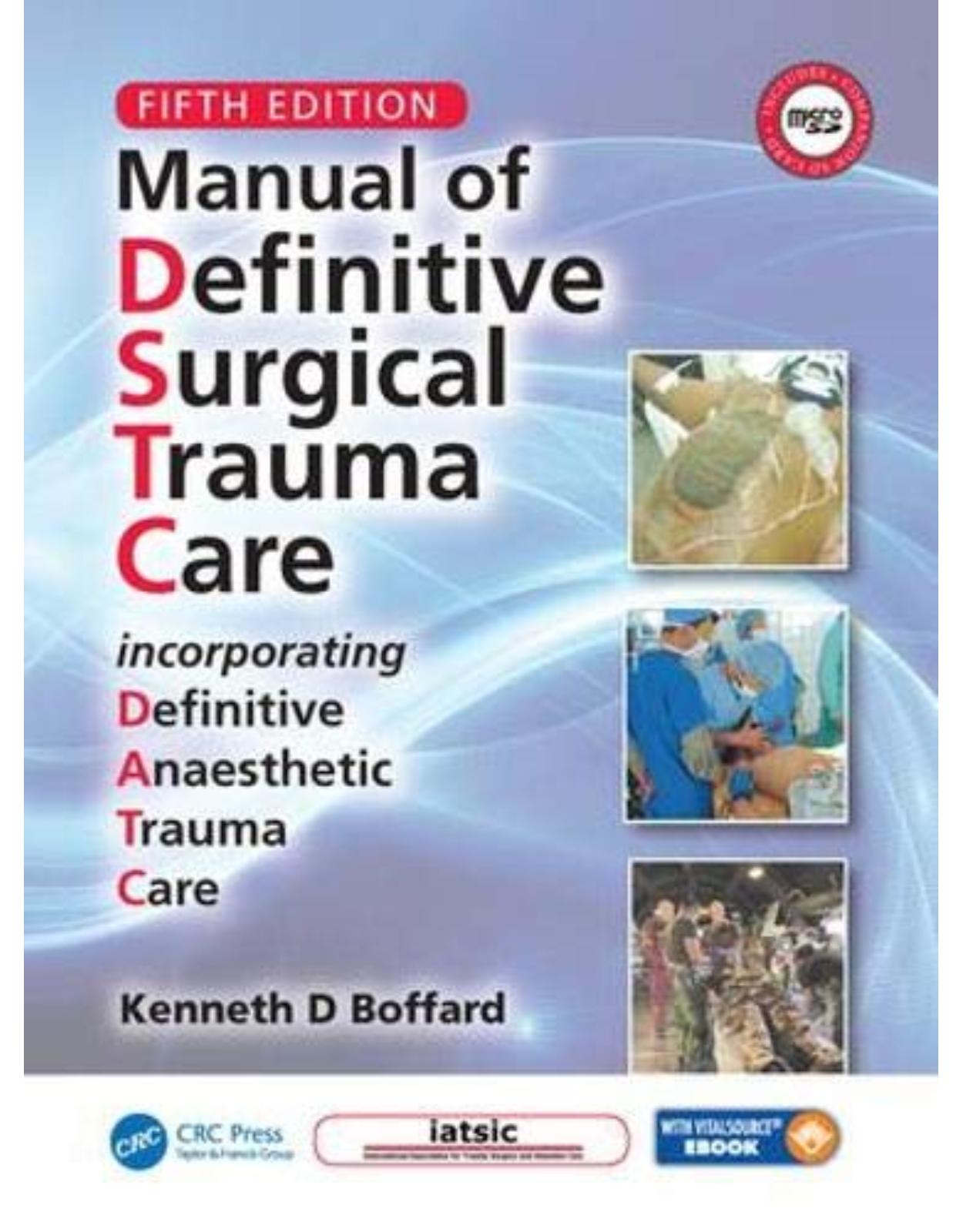
Manual of Definitive Surgical Trauma Care, Fifth Edition
Livrare gratis la comenzi peste 500 RON. Pentru celelalte comenzi livrarea este 20 RON.
Disponibilitate: La comanda in aproximativ 4 saptamani
Autor: Kenneth David Boffard
Editura: CRC Press
Limba: Engleza
Nr. pagini: 464
Coperta: Hardcover
Dimensiuni: 19.05 x 1.91 x 24.77 cm
An aparitie: 28 Jun. 2019
Description:
Developed for the International Association for Trauma Surgery and Intensive Care (IATSIC), the Manual of Definitive Surgical Trauma Care 5e is ideal for training all surgeons who encounter major surgical trauma on an infrequent basis. This new edition includes both an e-version, and also a microSD card containing over 20 operative videos. The increasing role of non-operative management (NOM) has been recognised, and the Military Module is substantially updated to reflect recent conflict experience. An expanded section highlights trauma management under austere conditions.Written by faculty who teach the DSTC Course, this definitive and well established book focuses on life-saving surgical techniques to use in challenging and unfamiliar incidents of trauma.
Table of Contents:
Video Contents
Preface
Introduction
Injury Prevention
Training in the Initial Management of Severe Trauma
The DSTC™ Course
The DATC™ Course
Summary
Board of Contributors
Acknowledgements
About the Author
Part 1: Trauma system and communication principles
1. Safe and Sustainable Trauma Care
1.1 Introduction
1.2 Safe Trauma Care
1.2.1 Individual Factors
1.2.1.1 Heuristics AND Cognitive Biases
1.2.1.2 Individual – Leadership
1.2.1.3 Trauma Team
1.2.1.4 Team – Training
1.2.2 Institutional Factors
1.2.2.1 Dedicated Trauma Service
1.2.3 Performance Improvement Activities
1.2.4 Regional Activities
1.2.5 National Activities
1.2.6 Global Activities
1.3 Sustainable Trauma Care
1.3.1 Workforce Development
1.4 Conclusion
References
2. Communication and Non-Technical Skills for Surgeons (NOTSS) in Major Trauma: The Role of Crew Resource Management (CRM)
2.1 Overview
2.1.1 The ‘Swiss Cheese’ Theory
2.2 Communication in the Trauma Setting
2.2.1 Initial Handover
2.2.2 Resuscitation and Ongoing Management
2.3 Leadership in Trauma Care
2.4 Potential Errors Related to Each Behavioural Theme
2.5 Summary
References and Recommended Reading
3. Pre-Hospital and Emergency Trauma Care
3.1 Resuscitation in the Emergency Department and Pre-hospital Setting
3.2 Management of Major Trauma
3.2.1 Resuscitation
3.2.1.1 Civilian Pre-hospital Tourniquet use
3.2.1.2 Primary Survey
3.2.1.3 Secondary Survey
3.2.2 Management of Penetrating Trauma
3.3 Emergency Department Surgery
3.3.1 Head Trauma
3.3.2 Chest Trauma
3.3.3 Abdominal Trauma
3.3.4 Pelvic Trauma
3.3.5 Long Bone Fractures
3.3.6 Peripheral Vascular Injuries
3.4 Summary
References and Recommended Reading
Part 2: Physiology and the body's response to trauma
4. Resuscitation Physiology
4.1 Metabolic Response to Trauma
4.1.1 Definition of Trauma
4.1.2 Initiating Factors
4.1.2.1 Hypovolaemia
4.1.2.2 Afferent Impulses
4.1.2.3 Wound Factors: Inflammatory and Cellular
4.1.2.4 Toxins/Sepsis
4.1.2.5 Free Radicals
4.1.2.6 Hypovolaemia
4.1.2.7 Afferent Impulses
4.1.2.8 Wound Factors
4.1.3 Immune Response
4.1.3.1 The Inflammatory Pathway
4.1.3.2 The Cellular Pathway
4.1.3.3 Toxins
4.1.3.4 PAMPS and DAMPS
4.1.3.5 Free Radicals
4.1.4 Hormonal Mediators
4.1.4.1 Hypothalamus/Pituitary
4.1.4.2 Adrenal Hormones
4.1.4.3 Pancreatic Hormones
4.1.4.4 Renal Hormones
4.1.4.5 Other Hormones
4.1.5 Effects of the Various Mediators
4.1.5.1 Hyperdynamic State
4.1.5.2 Water and Salt Retention
4.1.5.3 Effects on Substrate Metabolism
4.1.6 The Anabolic Phase
4.1.7 Clinical and Therapeutic Relevance
4.2 Shock
4.2.1 Definition of Shock
4.2.2 Classification of Shock
4.2.2.1 Hypovolaemic Shock
4.2.2.2 Cardiogenic Shock
4.2.2.3 Cardiac Compressive Shock
4.2.2.4 Distributive (Inflammatory) Shock
4.2.2.5 Neurogenic Shock
4.2.2.6 Obstructive Shock
4.2.3 Measurements in Shock
4.2.3.1 Cardiac Output
4.2.3.2 Indirect Measurement of Flow
4.2.3.3 Direct Measurements
4.2.4 Endpoints in Shock Resuscitation13
4.2.5 Post-Shock and Multiple Organ Failure Syndromes
4.2.6 Management of the Shocked Patient
4.2.6.1 Oxygenation
4.2.6.2 Fluid Therapy for Volume Expansion
4.2.6.3 Route of Administration
4.2.6.4 Pharmacologic Support of Blood Pressure
4.2.7 Prognosis in Shock
4.2.8 Recommended Protocol for Shock
4.2.8.1 Military Experience
4.2.8.2 Initial Resuscitation
REFERENCES AND RECOMMENDED READING
5. Transfusion in Trauma
5.1 Indications for Transfusion
5.1.1 Oxygen-Carrying Capacity
5.2 Transfusion Fluids
5.2.1 Colloids
5.2.1.1 Starches
5.2.1.2 Albumin
5.2.2 Blood
5.2.2.1 Fresh Whole Blood (FWB)
5.2.2.2 Packed red Blood Cells
5.2.3 Component Therapy (Platelets, Fresh Frozen Plasma, Cryoprecipitate)
5.2.3.1 Platelets
5.2.3.2 Fresh Frozen Plasma
5.2.3.3 Cryoprecipitate
5.2.3.4 Fibrinogen Concentrate
5.3 Effects of Transfusing Blood and Blood Products
5.3.1 Metabolic Effects
5.3.2 Effects of Microaggregates
5.3.3 Hyperkalaemia
5.3.4 Coagulation Abnormalities
5.3.5 Other Risks of Transfusion
5.3.5.1 Transfusion-transmitted Infections
5.3.5.2 Haemolytic Transfusion Reactions
5.3.5.3 Immunological Complications
5.3.5.4 Factors Implicated in Haemostatic Failure
5.4 Current Best Transfusion Practice
5.4.1 Initial Response
5.4.2 Reduction in the Need for Transfusion
5.4.3 Transfusion Thresholds
5.4.4 Transfusion Ratios
5.4.5 Adjuncts to Enhance Clotting
5.4.5.1 Recombinant Activated Factor VII (rFVIIa)
5.4.5.2 Tranexamic Acid (TXA)
5.4.5.3 Desmopressin (DDAVP)
5.4.6 Monitoring the Coagulation Status: Traditional and VHA
5.4.6.1 Traditional Assays
5.4.6.2 Viscoelastic Haemostatic Assays (VHA): Thromboelastography (TEG)/Rotary Thromboelastomerography (RoTEM)19,20
5.5 Autotransfusion
5.6 Red Blood Cell Substitutes23
5.6.1 Perfluorocarbons
5.6.2 Haemoglobin Solutions
5.6.2.1 Liposomal Haemoglobin Solutions
5.6.2.2 Polymerized Haemoglobin Solutions (Human-Outdated/Bovine RBCs)
5.6.3 Future Evolution
5.7 Massive Haemorrhage/Massive Transfusion24
5.7.1 Definition
5.7.2 Massive Transfusion Protocol (MTP)
5.8 Haemostatic Adjuncts in Trauma
5.8.1 Overview
5.8.2 Tissue Adhesives
5.8.2.1 Fibrin
5.8.2.2 Tachosil®
5.8.2.3 HemoPatch®
5.8.2.4 Collagen Fleece
5.8.3 Other Haemostatic Adjuncts
5.8.3.1 Chitosan (Celox (Meditrade Ltd., Crewe, UK)/HemCon (HemCon Medical Technologies, Portland, OR, USA))
5.8.3.2 Mineral Zeolyte (QuikClot® (Z-Medical Corporation, Wallingford CT, USA))
References and Recommended Reading
6. Damage Control
6.1 Introduction
6.2 Damage Control Resuscitation
6.3 Damage Control Surgery
6.3.1 Stage 1: Patient Selection
6.3.2 Stage 2: Operative Haemorrhage and Contamination Control
6.3.2.1 Initial Incision
6.3.2.2 Haemorrhage Control
6.3.2.3 Control of Contamination (Hollow Viscus Organs)
6.3.2.4 Copious Washout
6.3.2.5 Temporary Abdominal Closure (TAC)
6.3.3 Stage 3: Physiological Restoration in the ICU
6.3.3.1 Restoration of Body Temperature
6.3.3.2 Optimization of Oxygen Delivery
6.3.3.3 Correction of Clotting Profiles
6.3.3.4 Improvement of Physiological Endpoints: as Evidenced by
6.3.3.5 Monitoring for and Minimizing the Incidence of Intra-Abdominal Hypertension (AH) and Abdominal Compartment Syndrome (ACS) (See also Section 17.10)
6.3.3.6 Recognition of Additional Injuries
6.3.4 Stage 4: Definitive Surgery
6.3.4.1 The ‘Re-Look Laparotomy’
6.3.5 Stage 5: Abdominal Wall Closure
6.3.5.1 Delayed Primary Abdominal Closure
6.3.5.2 Secondary Abdominal Closure
6.3.5.3 Secondary Closure
6.3.5.4 Planned Hernia
6.3.6 Outcomes
6.4 Damage Control Orthopaedics17
References and recommended reading
Part 3: Anatomical and organ system injury
7. The Neck
7.1 Overview
7.2 Management Principles: Penetrating Cervical Injury
7.2.1 Initial Assessment and Definitive Airway
7.2.2 Control of Haemorrhage
7.2.3 Injury Location
7.2.4 Mechanism
7.2.5 Frequency of Injury
7.2.6 Use of Diagnostic Studies
7.2.6.1 Computed Tomography Scanning with Contrast/Computed Tomography Angiography
7.2.6.2 Angiography
7.2.6.3 Other Diagnostic Studies
7.3 Management
7.3.1 Mandatory versus Selective Neck Exploration
7.3.2 Management Based on Anatomical Zones
7.4 Access to the Neck
7.4.1 Position
7.4.2 Incision
7.4.3 Surgical Access
7.4.3.1 Zone I
7.4.3.2 Zone II
7.4.3.3 Zone III
7.4.4 Priorities
7.4.4.1 Vascular Injuries
7.4.4.2 Carotid Artery
7.4.4.3 Tracheal Injuries
7.4.4.4 Pharyngeal and Oesophageal Injuries
7.4.5 Midline Visceral Structures
7.4.6 Root of the Neck
7.4.7 Collar Incisions
7.4.8 Vertebral Arteries
References and recommended reading
8. The Chest
8.1 Overview
8.2 The Spectrum of Thoracic Injury
8.2.1 Immediately Life-Threatening Injuries
8.2.2 Potentially Life-Threatening Injuries
8.3 Pathophysiology of Thoracic Injuries
8.3.1 Paediatric Considerations
8.4 Applied Surgical Anatomy of the Chest
8.4.1 The Chest Wall
8.4.2 The Chest Floor
8.4.3 The Chest Contents
8.4.3.1 Tracheobronchial Tree
8.4.3.2 Lungs and Pleurae
8.4.3.3 Heart and Pericardium
8.4.3.4 The Aorta and Great Vessels
8.4.3.5 Oesophagus
8.4.3.6 Thoracic Duct
8.5 Diagnosis
8.6 Management of Specific Injuries
8.6.1 Damage Control in the Chest
8.6.2 Open Pneumothorax
8.6.3 Tension Pneumothorax (Haemo/Pneumothorax)
8.6.4 Massive Haemothorax
8.6.5 Tracheobronchial Injuries12
8.6.6 Oesophageal Injuries
8.6.7 Diaphragmatic Injuries
8.6.8 Pulmonary Contusion13
8.6.9 Flail Chest13
8.6.10 Fixation of Multiple Fractures of Ribs
8.6.11 Pulmonary Laceration
8.6.12 Air Embolism15
8.6.13 Cardiac Injuries
8.6.14 Injuries to the Great Vessels
8.7 Chest Drainage
8.7.1 Drain Insertion
8.7.2 Drain Removal
8.8 Surgical Approaches to the Thorax
8.8.1 Anterolateral Thoracotomy
8.8.1.1 Technique
8.8.1.2 Closure
8.8.2 Median Sternotomy
8.8.2.1 Technique
8.8.2.2 Closure
8.8.3 The ‘Clamshell’ Thoracotomy
8.8.4 Posterolateral Thoracotomy
8.8.5 ‘Trapdoor’ Thoracotomy
8.9 Emergency Department Thoracotomy
8.9.1 History
8.9.2 Objectives
8.9.3 Indications and Contraindications
8.9.4 Results
8.9.5 When to Stop EDT
8.9.6 Technique
8.9.6.1 Instrument Requirements
8.9.6.2 Approach
8.10 Surgical Procedures
8.10.1 Pericardial Tamponade
8.10.2 Cardiac Injury
8.10.3 Pulmonary Haemorrhage
8.10.4 Pulmonary Tractotomy
8.10.5 Lobectomy or Pneumonectomy22
8.10.6 Thoracotomy with Aortic Cross-Clamping
8.10.7 Aortic Injury
8.10.8 Tracheobronchial Injury
8.10.9 Oesophageal Injury
8.11 Summary
8.12 Anaesthesia for Thoracic Trauma
8.12.1 Penetrating Thoracic Injury
8.12.2 Blunt Thoracic Injury
8.12.2.1 Contained Large Vessel Rupture/Aneurism
8.12.2.2 Pulmonary Contusion
8.12.2.3 Large Airway Disruption
8.12.2.4 Flail Chest
8.12.2.5 Diaphragmatic Injury
8.12.3 Anaesthetic Management of Thoracic Injury
8.13 Anaesthetic Considerations
References and Recommended Reading
9. The Abdomen
9.1 The Trauma Laparotomy
9.1.1 Overview
9.1.1.1 Difficult Abdominal Injury Complexes
9.1.1.2 The Retroperitoneum
9.1.1.3 Non-Operative Management of Penetrating Abdominal Injury
9.1.2 The Trauma Laparotomy
9.1.2.1 Pre-Operative Adjuncts
9.1.2.2 Draping
9.1.2.3 Incision
9.1.2.4 Initial Procedure
9.1.2.5 Perform a Trauma Laparotomy
9.1.2.6 Perform Definitive Packing
9.1.2.7 Specific Routes of Access
9.1.2.8 Specific Organ Techniques (see also Specific Organs)
9.1.3 Closure of the Abdomen
9.1.3.1 Principles of Abdominal Closure
9.1.3.2 Choosing the Optimal Method of Closure
9.1.3.3 Primary Closure
9.1.4 Specific Tips and Tricks
9.1.4.1 Headlight
9.1.4.2 Stirrups and Lithotomy Position
9.1.4.3 Table Tilt
9.1.4.4 Be Flexible: Move!
9.1.4.5 Aortic Compression Spoon
9.1.4.6 Pericardial Window
9.1.4.7 Washout
9.1.4.8 Drains
9.1.4.9 Stomas
9.1.4.10 Temporary Closure
9.1.4.11 Two Catheters: Bladder Injury
9.1.4.12 Early Tracheostomy
9.1.5 Briefing for Operating Room Scrub Nurses
9.1.6 Summary
References and Further Reading
References
Recommended Reading
9.2 Abdominal Vascular Injury
9.2.1 Overview
9.2.2 Retroperitoneal Haematoma
9.2.2.1 Central Haematoma
9.2.2.2 Lateral Haematoma
9.2.2.3 Pelvic Haematoma
9.2.3 Surgical Approach to Major Abdominal Vessels
9.2.3.1 Incision
9.2.3.2 Medial Visceral Rotation (see also Section 9.1.2.7)
9.2.3.3 Coeliac Axis
9.2.3.4 Superior Mesenteric Artery3
9.2.3.5 Inferior Mesenteric Artery
9.2.3.6 Renal Arteries
9.2.3.7 Iliac Vessels
9.2.3.8 Inferior Vena Cava8
9.2.3.9 Portal Vein10
9.2.4 Shunting
References and Recommended Reading
References
Selected Reading
9.3 Bowel, Rectum, and Diaphragm
9.3.1 Overview
9.3.2 Diaphragm
9.3.3 Stomach
9.3.4 The Duodenum
9.3.5 Small Bowel
9.3.5.1 The Stable Patient
9.3.5.2 The Unstable Patient
9.3.6 Large Bowel3,4
9.3.6.1 The Stable Patient
9.3.6.2 The Unstable Patient
9.3.7 Rectum5
9.3.8 Mesentery
9.3.9 Adjuncts
9.3.9.1 Antibiotics
References and Selected Readings
References
Selected Readings
9.4 The Liver and Biliary System
9.4.1 Overview
9.4.2 Resuscitation
9.4.3 Diagnosis
9.4.4 Liver Injury Scale4
9.4.5 Management
9.4.5.1 Subcapsular Haematoma
9.4.5.2 Non-Operative Management (NOM)5,6
9.4.5.3 Subcapsular Haematoma
9.4.5.4 Operative (Surgical) Management
9.4.6 Surgical Approach8–10
9.4.6.1 Incision
9.4.6.2 Initial Actions
9.4.6.3 Techniques for Temporary Control of Haemorrhage
9.4.6.4 Mobilization of the Liver
9.4.6.5 Hepatic Isolation
9.4.7 Perihepatic Drainage
9.4.8 Complications
9.4.9 Injury to the Retrohepatic Vena Cava
9.4.10 Injury to the Porta Hepatis13
9.4.11 Injury to the Bile Ducts and Gallbladder14,15
9.4.12 Anaesthetic Considerations
References
Recommended Reading
9.5 Spleen
9.5.1 Overview
9.5.2 Anatomy
9.5.3 Diagnosis
9.5.3.1 Clinical
9.5.3.2 Ultrasound
9.5.3.3 Computed Tomography (CT) Scan
9.5.4 Splenic Injury Scale1
9.5.5 Management
9.5.5.1 Non-Operative Management2
9.5.5.2 Operative Management
9.5.6 Surgical Approach
9.5.6.1 Spleen Not Actively Bleeding
9.5.6.2 Splenic Surface Bleed Only
9.5.6.3 Minor Lacerations
9.5.6.4 Splenic Tears
9.5.6.5 Partial Splenectomy
9.5.6.6 Mesh Wrap
9.5.6.7 Splenectomy
9.5.6.8 Drainage
9.5.7 Outcome
9.5.8 Opportunistic Post-Splenectomy Infection
References and Recommended Reading
9.6 Pancreas
9.6.1 Overview
9.6.2 Anatomy
9.6.3 Mechanisms of Injury
9.6.3.1 Blunt Trauma
9.6.3.2 Penetrating Trauma
9.6.4 Diagnosis
9.6.4.1 Clinical Evaluation
9.6.4.2 Serum Amylase and Serum Lipase
9.6.4.3 Ultrasound
9.6.4.4 Diagnostic Peritoneal Lavage (DPL)
9.6.4.5 Computed Tomography
9.6.4.6 Endoscopic Retrograde Cholangiopancreatography
9.6.4.7 Magnetic Resonance Cholangiopancreatography
9.6.4.8 Intra-operative Pancreatography
9.6.4.9 Operative Evaluation
9.6.5 Pancreas Injury Scale
9.6.6 Management
9.6.6.1 Non-Operative Management
9.6.6.2 Operative Management
9.6.7 Surgical Approach
9.6.7.1 Incision and Exploration (see also SECTION 9.1)
9.6.7.2 Pancreatic Injury: Surgical Decision-Making
9.6.8 Adjuncts
9.6.8.1 Somatostatin and Its Analogues
9.6.8.2 Nutritional Support
9.6.9 Pancreatic Injury in Children
9.6.10 Complications
9.6.10.1 Early Complications
9.6.10.2 Late Complications
9.6.11 Summary of Evidence Based Guidelines31,32
References and Recommended Reading
9.7 The Duodenum
9.7.1 Overview
9.7.2 Mechanism of Injury
9.7.2.1 Penetrating Trauma
9.7.2.2 Blunt Trauma
9.7.2.3 Paediatric Considerations
9.7.3 Diagnosis
9.7.3.1 Clinical Presentation
9.7.3.2 Serum Amylase and Serum Lipase
9.7.3.3 Diagnostic Peritoneal Lavage/Ultrasound
9.7.3.4 Radiological Investigation
9.7.3.5 Diagnostic Laparoscopy
9.7.4 Duodenal Injury Scale
9.7.5 Management
9.7.6 Surgical Approach
9.7.6.1 Intramural Haematoma
9.7.6.2 Duodenal Laceration
9.7.6.3 Repair of the Perforation
9.7.6.4 Complete Transection of the Duodenum
9.7.6.5 Duodenal Diversion
9.7.6.6 Duodenal Diverticulation
9.7.6.7 Triple Tube Decompression14
9.7.6.8 Pyloric Exclusion (see also Section 9.6.7.2.7)
References and Recommended Reading
9.8 The Urogenital System
9.8.1 Overview
9.8.2 Renal Injuries
9.8.2.1 Diagnosis
9.8.2.2 Renal Injury Scale
9.8.2.3 Management
9.8.2.4 Surgical Approach
9.8.2.5 Adjuncts
9.8.2.6 Post-operative Care
9.8.3 Ureteric Injuries
9.8.3.1 Diagnosis
9.8.3.2 Surgical Approach
9.8.3.3 Complications
9.8.4 Bladder Injuries
9.8.4.1 Diagnosis
9.8.4.2 Management
9.8.4.3 Surgical Approach
9.8.5 Urethral Injuries
9.8.5.1 Diagnosis
9.8.5.2 Management
9.8.5.3 Ruptured Urethra
9.8.6 Injury to the Scrotum
9.8.6.1 Diagnosis
9.8.6.2 Management
9.8.7 Gynaecological Injury and Sexual Assault
9.8.7.1 Management
9.8.8 Injury of the Pregnant Uterus
References and Recommended Reading
10. The Pelvis
10.1 Anatomy
10.2 Classification
10.2.1 Tile’s Classification2
10.2.1.1 Type A: Completely stable
10.2.1.2 Type B: Vertically stable but rotationally unstable
10.2.1.3 Type C: Wholly unstable in rotational and vertical planes
10.2.1.4 A Jumper‘s Fracture
10.2.1.5 Acetabular Fractures
10.2.1.6 Fracture Combinations
10.2.2 Young and Burgess Classification3
10.2.2.1 Anteroposterior Compression (APC) (Type 1, 2, 3)
10.2.2.2 Lateral Compression (LC) (Type 1, 2, 3)
10.2.2.3 Vertical Shear
10.2.2.4 Combined Mechanism
10.3 Clinical Examination and Diagnosis
10.4 Resuscitation
10.4.1 Haemodynamically Normal Patients
10.4.2 Haemodynamically Stable Patients (Transient Responders)
10.4.3 Haemodynamically Unstable Patients (Non-Responders)
10.5 External Fixation
10.5.1 Iliac-Crest Route
10.5.2 Supra-acetabular Route
10.5.3 Pelvic C-clamp
10.6 Laparotomy
10.7 Extraperitoneal pelvic Packing
10.7.1 Technique of Extraperitoneal Packing11,12
10.8 Associated Injuries
10.8.1 Head Injuries
10.8.2 Intra-abdominal Injuries
10.8.3 Bladder and Urethral Injuries (See also Section 9.8)
10.8.4 Urethral Injuries (See Section 9.8)
10.8.5 Anorectal Injuries14
10.8.6 Vaginal Injuries
10.9 Open Pelvic Fractures
10.9.1 Diagnosis
10.9.2 Surgery
10.10 Summary
References and recommended reading
11. Extremity Trauma
11.1 Overview
11.2 Management of Severe Injury to the Extremity
11.3 Management of Vascular Injury of the Extremity
11.3.1 Chemical Vascular Injuries
11.4 Crush Syndrome
11.5 Management of Open Fractures
11.5.1 Severity of Injury (Gustilo Classification)2
11.5.2 Sepsis and Antibiotics
11.5.3 Venous Thromboembolism
11.5.4 Timing of Skeletal Fixation in Polytrauma Patients
11.5.4.1 Respiratory Insufficiency8
11.5.4.2 Head Injury
11.6 Massive Limb Trauma: life versus Limb
11.6.1 Scoring Systems
11.6.1.1 Mangled Extremity Syndrome Index (MESI)
11.6.1.2 Predictive Salvage Index System
11.6.1.3 Mangled Extremity Severity Score (MESS)
11.6.1.4 Nisssa Scoring System
11.7 Compartment Syndrome16–18
11.8 Fasciotomy
11.8.1 Lower Leg Fasciotomy
11.8.1.1 Two Incision–Four Compartment Fasciotomy20
11.8.1.2 Single Incision Fasciotomy
11.8.1.3 Fibulectomy
11.8.1.4 Subcutaneous Fasciotomy
11.8.2 Upper Leg21
11.8.3 Upper and Lower Arm22,23
11.9 Complications of Major Limb Injury
11.10 Summary
References and recommended reading
12. Head Trauma
12.1 Introduction
12.2 Injury Patterns and Classification
12.2.1 Severity
12.2.2 Pathological Classification of TBI
12.2.2.1 Blunt Head Trauma
12.2.2.2 Penetrating Head Trauma
12.3 Measurable physiological Parameters IN TBI
12.3.1 Mean Arterial Pressure
12.3.2 Intracranial Pressure
12.3.3 Cerebral Perfusion Pressure
12.3.4 Cerebral Blood Flow
12.4 Pathophysiology of Traumatic Brain Injury4
12.5 Management of TBI
12.6 Cerebral Perfusion Pressure Threshold
12.7 Intracranial pressure Monitoring and Threshold
12.7.1 ICP Monitoring Devices
12.7.1.1 CSF Drainage
12.7.2 ICP Management – Do’s and Don'ts
12.7.2.1 Hyperventilation
12.7.2.2 Osmotherapy (Mannitol and Hypertonic Saline)
12.7.2.3 Barbiturates and Propofol
12.7.2.4 Steroids
12.8 Imaging
12.9 Indications for Surgery
12.9.1 Burr Holes and Emergency Craniotomy
12.9.1.1 Emergency Burr Hole craniotomy9
12.9.1.2 Emergency Craniotomy
12.10 Adjuncts to Care
12.10.1 Infection Prophylaxis
12.10.2 Seizure Prophylaxis
12.10.3 Nutrition
12.10.4 Deep Vein Thrombosis Prophylaxis
12.10.5 Steroids
12.11 Paediatric Considerations
12.12 Pearls and Pitfalls
12.13 Summary
12.14 Anaesthetic Considerations
References and Recommended Reading
13. Burns
13.1 Overview
13.2 Burns Pathophysiology
13.3 Anatomy
13.4 Special Types of Burn
13.4.1 Chemical Burns
13.4.2 Electrical Injury
13.5 Depth of the Burn
13.5.1 Superficial Burn (Erythema)
13.5.2 Superficial Partial Thickness
13.5.3 Deep Partial Thickness
13.5.4 ‘Indeterminate’ Partial Thickness Burns
13.5.5 Full Thickness
13.6 Total Body Surface Area Burned
13.7 Management
13.7.1 Safe Retrieval
13.7.2 First Aid
13.7.3 Initial Management
13.7.3.1 Airway
13.7.3.2 Analgesia
13.7.3.3 Intravenous Access
13.7.3.4 Emergency Management of the Burn Wound
13.7.3.5 Fluid Resuscitation
13.7.3.6 Associated Injuries
13.7.4 Escharotomy and Fasciotomy
13.7.5 Definitive Management
13.7.5.1 ‘Closing’ the Burn Wound
13.7.5.2 Technique of Excision and Split Skin Grafting
13.7.5.3 Tumescent Technique
13.7.5.4 Wound Coverage
13.7.5.5 Burn Wound Excision and Closure
13.7.6 Assessing and Managing Airway Burns
13.7.6.1 Upper Airway
13.7.6.2 Lower Airway
13.7.6.3 Inhalational Toxicity
13.7.7 Tracheostomy
13.8 Special Areas
13.8.1 Face
13.8.2 Hands
13.8.3 Perineum
13.8.4 Feet
13.9 Adjuncts in Burn Care
13.9.1 Nutrition in the Burned Patient6
13.9.1.1 Paediatric Burn Nutrition
13.9.2 Ulcer Prophylaxis9 (See Table 17.2 S)
13.9.3 Venous Thromboembolism Prophylaxis7,10 (See Table 17.2 R)
13.9.4 Vitamin C
13.9.5 Antibiotics
13.9.6 Other Adjuncts
13.10 Summary
References and recommended reading
14. Special Patient Situations
14.1 Paediatric Trauma
14.1.1 Introduction
14.1.2 Injury Patterns
14.1.3 Pre-Hospital
14.1.4 Resuscitation Room
14.1.4.1 Airway
14.1.4.2 Ventilation
14.1.4.3 Circulation
14.1.4.4 Disability
14.1.4.5 Cardiac Arrest
14.1.5 Specific Organ Injury
14.1.5.1 Head Injury
14.1.5.2 Thoracic Injury
14.1.5.3 Abdominal Injury5
14.1.5.4 Genitourinary Injury
14.1.5.5 Pelvic Injury
14.1.5.6 Suspected non-Accidental Injury
14.1.6 Analgesia
14.2 Trauma in the Elderly
14.2.1 Definition of ‘Older’ and Susceptibility to Trauma
14.2.2 Access to Trauma Care
14.2.3 Physiology
14.2.3.1 Respiratory System
14.2.3.2 Cardiovascular System
14.2.3.3 Nervous System
14.2.3.4 Renal
14.2.3.5 Musculoskeletal
14.2.3.6 Influence of Comorbid Conditions
14.2.4 Multiple Medications – Polypharmacy
14.2.5 Analgesia
14.2.6 Decision to Operate
14.2.7 Anaesthetic Considerations in the Elderly13
14.3 Trauma in Pregnancy14
14.3.1 Evaluation
14.4 Non-Beneficial (Futile) Care
References and Recommended reading
Part 4: Modern therapeutic and diagnostic technology
15. Minimal Access Surgery in Trauma
15.1 Laparoscopy
15.1.1 Screening Laparoscopy
15.1.2 Diagnostic Laparoscopy
15.1.3 Non-Therapeutic Laparoscopy
15.1.4 Therapeutic Laparoscopy
15.1.5 Technique
15.1.6 Risks
15.1.7 Applications
15.1.7.1 Bowel injury
15.1.7.2 Splenic injury
15.1.7.3 Liver injury
15.1.7.4 After non-operative management
15.1.7.5 Diaphragmatic injury
15.2 Video-Assisted Thoracoscopic Surgery7
15.2.1 Technique
15.2.2 Applications
15.2.3 Summary
15.3 Resuscitative Endovascular Balloon Occlusion of the Aorta (REBOA)
15.3.1 Anatomy
15.3.2 Physiology
15.3.3 Insertion Technique
15.3.4 Monitoring
15.3.5 Total, Partial, and Intermittent Occlusion, and Targeted Blood Pressure
15.3.6 Perioperative and Post-operative Care
15.3.7 Indications
15.3.8 Contraindications
15.3.9 Complications16,17
15.3.10 Summary
15.4 Anaesthetic Considerations
References and Recommended Reading
16. Imaging in Trauma
16.1 Introduction
16.2 Radiation Doses and Protection from Radiation
16.3 Principles of Trauma Imaging
16.4 Pitfalls and Pearls
16.5 Trauma Ultrasound
16.5.1 Extended Focused Assessment by Sonography for Trauma
16.5.2 Indications and Results
16.5.2.1 Penetrating Abdominal Trauma
16.5.2.2 Blunt Abdominal Trauma
16.5.2.3 Pelvic Trauma
16.5.2.4 Blunt Thoracic Trauma
16.5.2.5 Penetrating Thoracic Trauma
16.5.3 Other Applications of Ultrasound in Trauma
16.5.4 Training
16.5.5 Summary
References and Recommended Reading
Part 5: Specialised aspects of total trauma care
17. Critical Care of the Trauma Patient
17.1 Introduction
17.2 Phases of ICU Care
17.2.1 Resuscitative Phase (First 24 Hours Post-Injury)1
17.2.1.1 ‘Traditional’ End Points of Resuscitation
17.2.1.2 Post-Traumatic Acute Lung Injury
17.2.1.3 Respiratory Assessment and Monitoring
17.2.2 Early Life Support Phase (24–72 Hours Post-Injury)
17.2.2.1 Priorities
17.2.3 Prolonged Life Support (>72 Hours Post-Injury)
17.2.3.1 Respiratory Failure
17.2.3.2 Infectious Complications
17.2.3.3 Non-Infectious Causes of Fever
17.2.3.4 Percutaneous tracheostomy6
17.2.3.5 Weaning from Ventilatory Support
17.2.3.6 Extubation Criteria (‘SOA2P’)
17.2.4 Recovery Phase (Separation from the ICU)
17.3 ExtraCorporeal Membrane Oxygenation8–10
17.3.1 Overview
17.3.2 Modes of ECMO
17.3.2.1 Veno-Venous ECMO (VV-ECMO)
17.3.2.2 Veno-Arterial ECMO (VA-ECMO)
17.3.2.3 Arterio-Venous ECMO (AV-ECMO)
17.3.3 Exclusions
17.4 Coagulopathy of Major Trauma11–13 (See also Chapter 5)
17.4.1 Management
17.5 Hypothermia
17.6 Multisystem Organ Dysfunction Syndrome (MODS)
17.7 Systemic Inflammatory Response Syndrome (See also Chapter 4)
17.8 Sepsis
17.8.1 Definitions
17.8.1.1 Sepsis
17.8.1.2 Severe Sepsis
17.8.1.3 Septic Shock
17.8.2 Surviving Sepsis Guidelines
17.9 Antibiotics
17.10 Abdominal Compartment Syndrome (ACS)
17.10.1 Introduction
17.10.2 Definition of ACS
17.10.3 Pathophysiology
17.10.4 Effect of Raised IAP on Individual Organ Function
17.10.4.1 Cardiovascular
17.10.4.2 Respiratory
17.10.4.3 Visceral Perfusion
17.10.4.4 Renal
17.10.4.5 Intracranial Pressure
17.10.5 Measurement of IAP
17.10.5.1 Measurement of APP
17.10.6 Management
17.10.6.1 Prevention
17.10.6.2 Treatment
17.10.6.3 Reversible Factors
17.10.7 Surgery for Raised IAP
17.10.7.1 Tips for Surgical Decompression for Raised IAP
17.10.8 Management Algorithm
17.11 Acute Kidney Injury37
17.12 Metabolic Disturbances
17.13 Nutritional Support39,40
17.13.1 Access for Enteral Nutrition
17.13.1.1 Simple
17.13.1.2 More Complicated
17.14 Prophylaxis in the ICU
17.14.1 Stress Ulceration44
17.14.2 Deep Venous Thrombosis and Pulmonary Embolus45
17.14.3 Tetanus Prophylaxis
17.14.4 Line Sepsis
17.15 Pain Control
17.16 ICU Tertiary Survey50
17.16.1 Evaluation for Occult Injuries
17.16.2 Assess Co-Morbid Conditions
17.16.3 ICU Summary
17.17 Family Contact and Support (See also Chapter 19)
References and Recommended Reading
18. Trauma Anaesthesia
18.1 Introduction
18.2 Planning and Communicating
18.3 Damage Control Resuscitation1
18.3.1 Limited Fluid Administration
18.3.2 Targeting Coagulopathy
18.3.3 Prevent and Treat Hypothermia
18.4 Damage Control Surgery
18.4.1 Anaesthetic Procedures
18.4.1.1 Airway
18.4.1.2 Breathing
18.4.1.3 Circulation
18.4.1.4 Vascular access
18.4.2 Monitoring
18.5 Anaesthesia Induction in Hypovolaemic Shock
18.5.1 Introduction
18.5.2 Drugs for Anaesthesia Induction
18.5.2.1 Propofol
18.5.2.2 Ketamine
18.5.2.3 Etomidate
18.5.2.4 Thiopental
18.5.2.5 Midazolam
18.6 Battlefield Anaesthesia (See also Chapters 21 and 22)
18.6.1 Damage Control Anaesthesia in the Military Setting
18.6.2 Battlefield Analgesia
References
19. Psychology of Trauma
19.1 What is Psychological Trauma?
19.2 Reactions to Trauma
19.3 Post-Traumatic Stress Disorder
19.4 Trauma and ICU
19.5 The Clinical Psychologist
19.5.1 The Role of the Clinical Psychologist
19.5.1.1 For the Patient
19.5.1.2 For the Family
19.5.1.3 For the Team
19.5.2 When to Call the Clinical Psychologist
Recommended Reading
20. Physical and Rehabilitation Medicine P&RM
20.1 Definition
20.2 The Rehabilitation ‘Team’
20.3 Rehabilitation Starts in ICU
20.4 Outcomes-Based Rehabilitation (OBR)
20.4.1 FIM/FAM Assessment2,3
20.4.2 Glasgow Outcome Scale4
20.4.3 Rancho Los Amigos Scale5
20.5 Summary
References and Recommended Reading
21. Austere Environments
21.1 Definition
21.2 Overview
21.3 Infrastructure
21.3.1 Location
21.3.2 Hospital Structures
21.3.2.1 Water Supply
21.3.2.2 Energy
21.3.2.3 Waste Disposal
21.3.2.4 Sterilization Department
21.3.2.5 Surgical Equipment
21.3.2.6 Blood Bank
21.3.3 Health Protection of the Deployed Surgical Team
21.3.3.1 Vector-borne Disease
21.3.3.2 Enteric Illness
21.3.3.3 Road Trauma
21.3.3.4 Physical, Sexual, and Mental Health
21.4 Surgical Techniques to Have in Mind
21.4.1 Bleeding Control
21.4.2 Control of Contamination
21.4.3 Treatment of War Wounds
21.4.4 Amputations
21.4.5 Stabilization of Fractures
21.4.6 Obstetrics
21.4.7 Anaesthesia2,3
21.5 Post-operative Care and Documentation
21.6 Summary
References and RECOMMENDED Reading
22. Military Environments
22.1 Introduction
22.2 Injury Patterns
22.3 Emergency Medical Services Systems
22.3.1 The Echelons of Medical Care
22.3.1.1 Role 1
22.3.1.2 Role 2
22.3.1.3 Role 2 Light Manoeuvre
22.3.1.4 Role 2 Enhanced
22.3.1.5 Role 3
22.3.1.6 Role 4
22.3.2 Incident Management and Multiple Casualties
22.3.2.1 Confirm
22.3.2.2 Clear
22.3.2.3 Cordon
22.3.2.4 Control
22.3.2.5 Command and Control
22.3.2.6 Safety
22.3.2.7 Communication
22.3.2.8 Assessment
22.3.2.9 Triage
22.3.2.10 Treatment
22.3.2.11 Transport
22.4 Triage
22.4.1 Source and Aim of Triage
22.4.2 Forward Surgical Teams and Triage
22.4.3 Forward Surgical Team Decision-Making
22.4.4 Selection of Patients for Surgery
22.5 Mass Casualties
22.6 Evacuation4,5
22.7 Resuscitation
22.7.1 Overview
22.7.2 Damage Control Resuscitation8
22.7.3 Damage Control Surgery in the Military Setting10–12
22.8 Blast Injury
22.8.1 Diagnosis and Management of Blast Injuries
22.8.1.1 Rupture of the Tympanic Membrane
22.8.1.2 Blast Lung Injury (BLI)
22.8.1.3 Intra-Abdominal Injuries
22.8.1.4 Other Injuries
22.9 Battlefield Analgesia13,14
22.10 Battlefield Anaesthesia
22.10.1 Induction of Anaesthesia
22.10.2 Maintenance of Anaesthesia
22.11 Critical Care (See also Chapter 17)
22.12 Translating Military Experience to Civilian Trauma Care15–17
22.12.1 Leadership
22.12.2 Front-End Processes
22.12.3 Common Training
22.12.4 Governance
22.12.5 Rehabilitation Services
22.12.6 Translational Research
22.13 Summary
References and Recommended Reading
Appendix A: Trauma Systems
A.1 Introduction
A.2 The Inclusive Trauma System
A.3 Components of an Inclusive Trauma System
A.3.1 Administration
A.3.2 Prevention
A.3.3 Public Education
A.4 Management of the Injured Patient within a System
A.5 Steps in Organizing a System
A.5.1 Public Support
A.5.2 Legal Authority
A.5.3 Establish Criteria for Optimal Care
A.5.4 Designation of Trauma Centres
A.5.5 System Evaluation
A.6 Results and Studies
A.6.1 Panel Review
A.6.2 Registry Study
A.6.3 Population-Based Studies
A.7 Summary
RECOMMENDED READING
Appendix B: Trauma Scores and Scoring Systems
B.1 Introduction
B.2 Physiological Scoring Systems
B.2.1 Glasgow Coma Scale1
B.2.2 Paediatric Trauma Score2
B.2.3 Revised Trauma Score3
B.2.4 Acute Physiologic and Chronic Health Evaluation II4
B.3 Anatomical Scoring Systems
B.3.1 Abbreviated Injury Scale6
B.3.2 The Injury Severity Score7
B.3.3 The New Injury Severity Score8
B.3.4 Anatomic Profile Score10
B.3.5 ICD-based Injury Severity Score11
B.3.6 Organ Injury Scaling System12
B.3.7 Penetrating Abdominal Trauma Index13
B.3.8 Revised Injury Severity Classification II14,15
B.4 Comorbidity Scoring Systems
B.5 Outcome Analysis
B.5.1 Functional Independence Measure and Functional Assessment Measure18,19
B.5.2 Glasgow Outcome Scale20
B.5.3 Major Trauma Outcome Study
B.5.4 A Severity Characterization of Trauma22,23
B.6 Comparison of Trauma Scoring Systems
B.7 Scaling System for Organ Specific Injuries12,26–32
B.8 Summary
REFERENCES
Appendix C: The Definitive Surgical Trauma Care Course: The Definitive Anaesthetic Trauma Care Course: Course Requirements And Syllabus
C.1 Background
C.2 Course Development and Testing
C.3 Course Details
C.3.1 Ownership
C.3.2 Mission Statement
C.3.3 Application to Hold a Course
C.3.4 Eligibility to Present
C.3.4.1 Local Organizations
C.3.4.2 National Organizations
C.3.5 Course Materials and Overview
C.3.6 Course Director
C.3.7 Course Faculty
C.3.8 Course Participants
C.3.9 Practical Skill Stations
C.3.10 Course Syllabus
C.3.11 Course Certification
C.4 Iatsic Recognition
C.5 Course Information
Appendix D: Definitive Surgical Trauma CareTM Course – Core Surgical Skills
D.1 The Neck
D.2 The Chest
D.3 The Abdominal Cavity
D.4 The Liver
D.5 The Spleen
D.6 The Pancreas
D.7 The Duodenum
D.8 The Genitourinary System
D.9 Abdominal Vascular Injuries
D.10 Peripheral Vascular Injuries
D.11 Insertion of Resuscitative Balloon Occlusion of the Aorta (REBOA) Catheter
Appendix E: Briefing for Operating Room Scrub Nurses
E.1 Introduction
E.2 Preparing the Operating Room
E.2.1 Environment
E.2.2 Blood Loss
E.2.3 Instruments
E.2.4 Cleaning
E.2.5 Draping
E.2.6 Adjuncts
E.3 Surgical Procedure
E.3.1 Instruments
E.3.2 Special Instruments and Improvised Gadgets
E.4 Abdominal Closure
E.5 Instrument and Swab Count
E.6 Medico-legal Aspects and Communication Skills
E.7 Critical Incident Stress Issues
E.8 Conclusion
References and recommended reading
Index
| An aparitie | 28 Jun. 2019 |
| Autor | Kenneth David Boffard |
| Dimensiuni | 19.05 x 1.91 x 24.77 cm |
| Editura | CRC Press |
| Format | Hardcover |
| ISBN | 9780367244682 |
| Limba | Engleza |
| Nr pag | 464 |

Clientii ebookshop.ro nu au adaugat inca opinii pentru acest produs. Fii primul care adauga o parere, folosind formularul de mai jos.