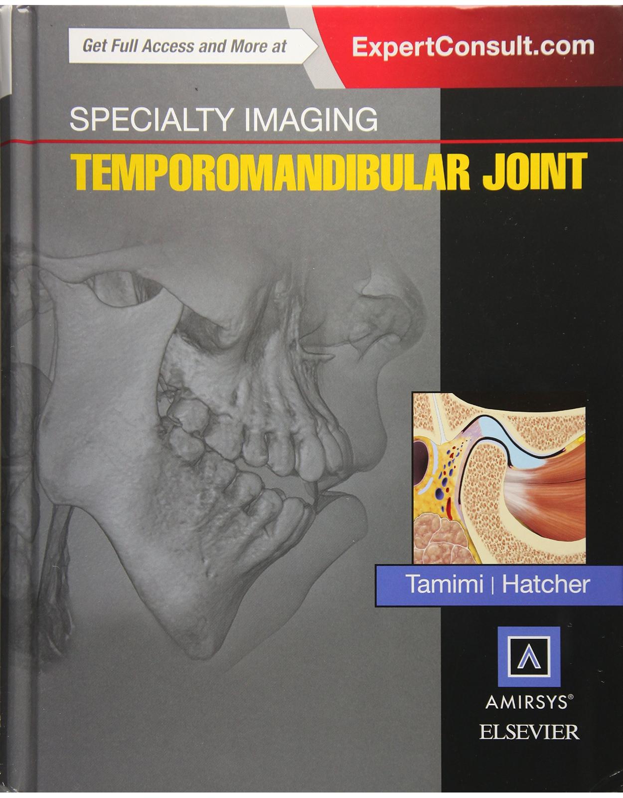
Specialty Imaging: Temporomandibular Joint
Produs indisponibil momentan. Pentru comenzi va rugam trimiteti mail la adresa depozit2@prior.ro sau contactati-ne la numarul de telefon 021 210 89 28 Vedeti mai jos alte produse similare disponibile.
Disponibilitate: Acest produs nu este momentan in stoc
Autor: Tamimi, Hatcher
Editura: Elsevier
Limba: Engleza
Nr. pagini: 800
Coperta: Hardcover
Dimensiuni: 22.9 x 5.1 x 28.6 cm
An aparitie: 2016
Description:
Specialty Imaging: Temporomandibular Joint offers expert insight into modern imaging of the temporomandibular joint by employing a multifaceted, multispecialty viewpoint of this difficult to understand joint. Image-rich content combines with easy-to-read text, bringing together the clinical perspectives and imaging expertise of today's research specialists.
- Includes extensive, in-depth explanations of the underlying mechanisms of normal vs. abnormal temporomandibular joints and how those present on radiographic imaging.
- Provides coverage of hot topics such as understanding the temporomandibular joint through biomechanical engineering, structure/function of the temporomandibular joint in normal and pathologic joints, and clinicoradiological correlation of temporomandibular joint findings.
- Details anatomic and functional interrelationships in conjunction with radiology.
Table of Contents
· Copyright
· DEDICATIONS
· CONTRIBUTING AUTHORS
· PREFACE
· ACKNOWLEDGMENTS
· SECTIONS
· SECTION 1: UNDERSTANDING THE TMJ
· Chapter 1: Embryology and Fetal Development of the Face and Neck
· Chapter 2: TMJ Embryology
· Chapter 3: TMJ Effect on Facial Growth
· Chapter 4: TMJ Effect on Upper Airway Morphology
· Chapter 5: Occlusion and Orthopedic Stability
· Chapter 6: Condylar Motions Related to Jaw Function
· Chapter 7: TMJ Biomechanics and Structure of the Mandibular Condyle
· Chapter 8: Structure and Function of the TMJ Disc and Disc Attachments
· Chapter 9: Modeling and Remodeling of TMJ and Mandible
· Regressive Remodeling
· Bone Modeling
· Bone Modeling
· Bone Modeling
· Bone Modeling
· SECTION 2: ANATOMY
· Chapter 10: TMJ Osseous Components
· Chapter 11: TMJ Disc/Fibrocartilage
· Chapter 12: TMJ Capsule and Ligaments
· Chapter 13: TMJ Histology and Synovial Fluid Composition
· Chapter 14: TMJ Innervation
· Chapter 15: TMJ Vasculature
· Chapter 16: Muscles of Mastication
· Chapter 17: Muscles of Facial Expression
· Chapter 18: Suprahyoid and Infrahyoid Neck
· Chapter 19: Tongue
· Chapter 20: Posterior Cervical Muscles
· Chapter 21: Mandible
· Chapter 22: Maxilla
· Chapter 23: Teeth
· Chapter 24: Temporal Bone
· Chapter 25: Intratemporal Facial Nerve
· Chapter 26: Skull Base Overview
· Chapter 27: Anterior Skull Base
· Chapter 28: Central Skull Base
· Chapter 29: Posterior Skull Base
· Chapter 30: Cranial Nerves Overview
· Chapter 31: Trigeminal Nerve (CNV)
· Chapter 32: Facial Nerve (CNVII)
· Chapter 33: Glossopharyngeal Nerve (CNIX)
· Chapter 34: Vagus Nerve (CNX)
· Chapter 35: Accessory Nerve (CNXI)
· Chapter 36: Hypoglossal Nerve (CNXII)
· Chapter 37: Cervical Spine
· Chapter 38: Craniocervical Junction
· Chapter 39: Styloid Process and Stylohyoid Ligament
· Chapter 40: Hyoid Bone
· Chapter 41: Sinonasal Overview
· Chapter 42: Nasopharynx and Oropharynx
· SECTION 3: MODALITIES USED FOR TMJ IMAGING
· Chapter 43: Imaging Decision Making
· Chapter 44: Plain Film Imaging
· Chapter 45: Arthrography
· Chapter 46: Introduction to CBCT Imaging
· Chapter 47: CBCT Analysis of TMJ
· Chapter 48: Radiation Dose in CBCT
· Chapter 49: Introduction to MDCT Imaging
· Chapter 50: Introduction to MR Imaging
· Chapter 51: Quantitative MR of Cartilage and Implications for TMJ Imaging
· Chapter 52: Arthroscopy
· SECTION 4: DIAGNOSES
· Chapter 53: Functional Disorders of Muscles
· Chapter 54: Intracapsular Disorders of TMJ
· Chapter 55: Correlation of Clinical Symptoms to Radiographic Findings
· Chapter 56: Craniofacial Malformations and Syndromes Affecting the TMJ
· Chapter 57: Hemifacial Microsomia
· Chapter 58: Treacher Collins Syndrome
· Chapter 59: Pierre Robin Sequence
· Chapter 60: Condylar Hypoplasia
· Chapter 61: Condylar Hyperplasia
· Chapter 62: Coronoid Hyperplasia
· Chapter 63: Hemimandibular Elongation
· Chapter 64: Mandibular Salivary Gland Defect (Stafne)
· Chapter 65: Foramen Tympanicum
· Chapter 66: TMJ Fracture, Adult and Neonatal
· Chapter 67: TMJ Dislocation
· Chapter 68: Bifid Condyle
· Chapter 69: Osteochondritis Dissecans
· Chapter 70: Rheumatoid Arthritis
· Chapter 71: Juvenile Idiopathic Arthritis
· Chapter 72: Pigmented Villonodular Synovitis
· Chapter 73: Chronic Recurrent Multifocal Osteomyelitis
· Chapter 74: Degenerative Joint Disease
· Chapter 75: Progressive Condylar Resorption
· Chapter 76: Synovial Cyst
· Chapter 77: MR Analysis of Normal TMJ Disc
· Chapter 78: Fine Structural Details of Disc and Posterior Attachment
· Chapter 79: Overview of Disc Displacements
· Chapter 80: Disc Displacement With Reduction
· Chapter 81: Disc Displacement Without Reduction
· Chapter 82: Joint Fluid and Marrow Alterations
· Chapter 83: Adhesions
· Chapter 84: Dual Bite
· Chapter 85: Fibrous Ankylosis
· Chapter 86: Bony Ankylosis
· Chapter 87: Osteoradionecrosis
· Chapter 88: Primary Synovial Chondromatosis
· Chapter 89: Secondary Synovial Chondromatosis
· Chapter 90: Calcium Pyrophosphate Dihydrate Deposition
· Chapter 91: Osteochondroma
· Chapter 92: Osteoma
· Chapter 93: Chondrosarcoma
· Chapter 94: Osteosarcoma
· Chapter 95: Metastasis
· Chapter 96: Simple Bone Cyst
· Chapter 97: Aneurysmal Bone Cyst
· Chapter 98: Fibrous Dysplasia
· Chapter 99: Attrition
· Chapter 100: Abfraction
· Chapter 101: Hypercementosis
· Chapter 102: Alveolar Process Exostosis
· Chapter 103: Torus Mandibularis
· Chapter 104: Torus Palatinus
· Chapter 105: Periapical Rarefying Osteitis
· Chapter 106: Oral Cavity Soft Tissue Infections
· Chapter 107: Osteomyelitis of the Jaw
· Chapter 108: Perineural Tumor Spread
· Chapter 109: Temporal Bone and Cervical Disorders Mimicking TMD
· Chapter 110: Temporal Bone Anatomy and Imaging Issues
· Chapter 111: EAC Atresia
· Chapter 112: Necrotizing External Otitis
· Chapter 113: EAC Cholesteatoma
· Chapter 114: EAC Keratosis Obturans
· Chapter 115: EAC Medial Canal Fibrosis
· Chapter 116: EAC Osteoma
· Chapter 117: EAC Skin SCCa
· Chapter 118: EAC Basal Cell Carcinoma
· Chapter 119: Acute Otomastoiditis With Coalescent Otomastoiditis
· Chapter 120: Acute Otomastoiditis With Abscess
· Chapter 121: Pars Flaccida Cholesteatoma
· Chapter 122: Labyrinthitis
· Chapter 123: Temporal Bone Fractures
· Chapter 124: Temporal Bone Fibrous Dysplasia
· Chapter 125: Temporal Bone Perineural Parotid Malignancy
· Chapter 126: Postradiated Temporal Bone
· Chapter 127: Degenerative Arthritis of CVJ
· Chapter 128: Cervical Facet Arthropathy
· Chapter 129: Cervical Spondylosis
· Chapter 130: Ankylosing Spondylitis, Cervical Spine
· Chapter 131: Adult Rheumatoid Arthritis, Cervical Spine
· Chapter 132: Rheumatoid Arthritis, Cervical Spine
· Chapter 133: Juvenile Idiopathic Arthritis, Cervical Spine
· Chapter 134: Ligamentous Injury
· Chapter 135: Ossification of Posterior Longitudinal Ligament
· Chapter 136: Diffuse Idiopathic Skeletal Hyperostosis
· Chapter 137: Calcium Pyrophosphate Dihydrate Deposition, Cervical Spine
· Chapter 138: Longus Colli Calcific Tendinitis
· Chapter 139: Masticator Space Overview
· Chapter 140: Pterygoid Venous Plexus Asymmetry
· Chapter 141: Benign Masticatory Muscle Hypertrophy
· Chapter 142: CNV3 Motor Denervation
· Chapter 143: Masticator Space Abscess
· Chapter 144: Masticator Space CNV3 Schwannoma
· Chapter 145: Masticator Space CNV3 Perineural Tumor
· Chapter 146: Masticator Space Chondrosarcoma
· Chapter 147: Masticator Space Sarcoma
· Chapter 148: Bell Palsy
· Chapter 149: Hemifacial Spasm
· Chapter 150: Trigeminal Neuralgia
· Chapter 151: Anterior Condylar Position
· Chapter 152: Posterior Condylar Position
· Chapter 153: Superior Condylar Position
· Chapter 154: Inferior Condylar Position
· Chapter 155: Small Condyle
· Chapter 156: Large Condyle
· Chapter 157: Large Coronoid Process
· Chapter 158: Well-Defined Erosion
· Chapter 159: Poorly Defined Erosion
· Chapter 160: TMJ Radiolucencies
· Chapter 161: TMJ Radiopacities
· Chapter 162: TMJ Soft Tissue Calcifications
· Chapter 163: Soft Tissue Calcifications, Head and Neck
· SECTION 6: CLINICAL DIFFERENTIAL DIAGNOSIS
· Chapter 164: Limited Oral Opening
· Chapter 165: Hypermobility
· Chapter 166: Joint Sounds
· Chapter 167: Tinnitus
· Chapter 168: Asymmetry
· Chapter 169: Posterior Open Bite
· Chapter 170: Anterior Open Bite
· SECTION 7: IMAGING OF TMJ PROCEDURES
· Chapter 171: TMJ Injection
· Chapter 172: Trigeminal Nerve Injection
· Chapter 173: Total Joint Replacement
· Chapter 174: TMJ Disc Replacement
· Chapter 175: TMJ Arthroscopic Surgery Cascade
| An aparitie | 2016 |
| Autor | Tamimi, Hatcher |
| Dimensiuni | 22.9 x 5.1 x 28.6 cm |
| Editura | Elsevier |
| Format | Hardcover |
| ISBN | 9780323377041 |
| Limba | Engleza |
| Nr pag | 800 |





Clientii ebookshop.ro nu au adaugat inca opinii pentru acest produs. Fii primul care adauga o parere, folosind formularul de mai jos.