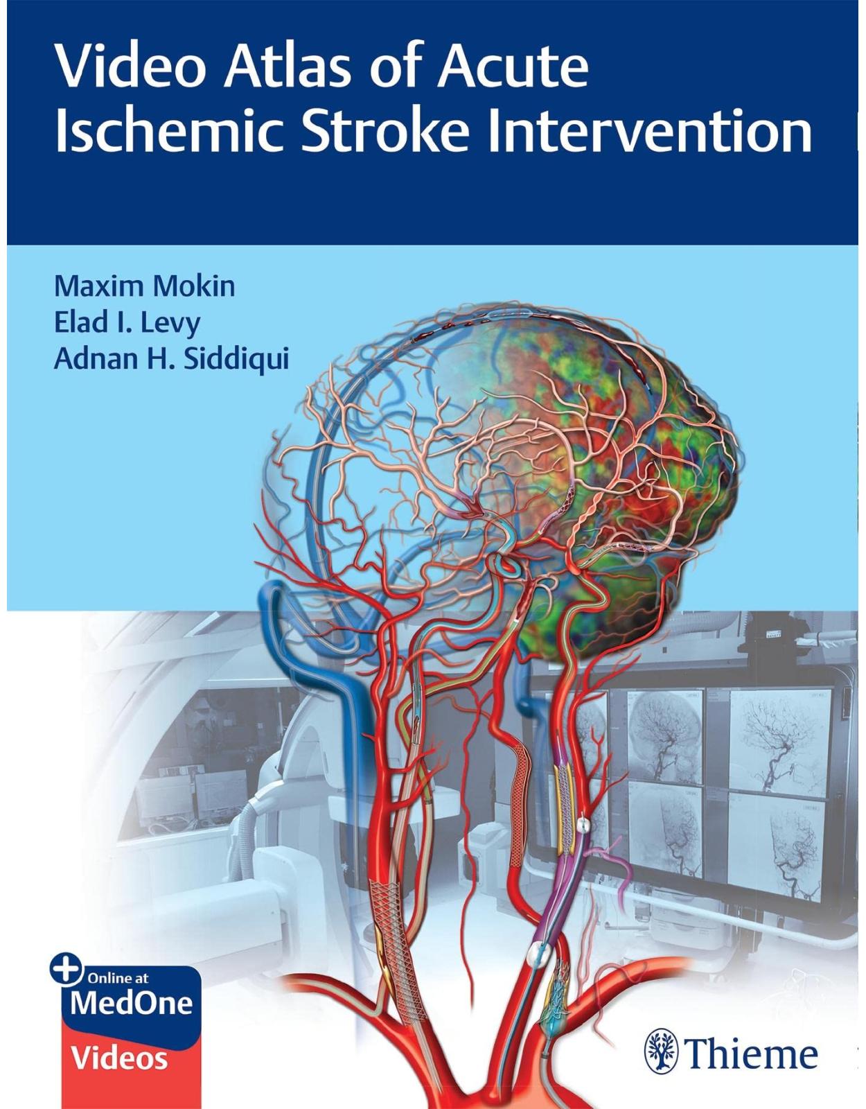
Video Atlas of Acute Ischemic Stroke Intervention
Livrare gratis la comenzi peste 500 RON. Pentru celelalte comenzi livrarea este 20 RON.
Disponibilitate: La comanda in aproximativ 4 saptamani
Editura: Thieme
Limba: Engleza
Nr. pagini: 212
Coperta:
Dimensiuni: 219 x 282 x 18 mm
An aparitie: 09 Mar 2022
Description:
The go-to resource for managing a full spectrum of clinical scenarios in acute ischemic stroke
Ever-evolving technological advances have created a daunting number of emergent neurointerventional protocols and therapies for treating acute ischemic stroke. This has created an urgent need for reader-friendly, hands-on resources. Video Atlas of Acute Ischemic Stroke Intervention edited by renowned neuroinventional stroke experts Maxim Mokin, Elad I. Levy, Adnan H. Siddiqui, and esteemed contributors fills a void in education and knowledge with interactive, case-based guidance on mastering challenging stroke interventions. The atlas leverages current knowledge and decades of experience in acute stroke treatment and device innovations to familiarize clinicians at various career stages with a wide repertoire of techniques available in the neurointerventional suite.
The book focuses on technical aspects of interventional stroke procedures including pitfalls and complications, an overview of the most useful tools of the trade, and discussion of pre- and post-procedural care. Eighteen chapters provide comprehensive coverage on the most common endovascular treatment approaches for acute stroke—aspiration and stent-retriever thrombectomy of proximal, distal, and tandem occlusions. Several cases focus on recognition and management of complications that clinicians may encounter during emergent procedures. Topics include the importance of arterial access, unmet need in current devices, and methods for overcoming these challenges.
Key Highlights
Thirty-eight individually narrated, high-definition videos describe angiographic and procedural cases, step by step
High-quality illustrations emphasize and delineate key aspects of complex procedures
Firsthand tips and tricks enhance the ability to manage highly challenging and less common pathologies, including arterial dissection, atherosclerosis, and venous strokes
This must-have resource will benefit all practitioners involved in the interventional care of patients with acute ischemic stroke.
Table of Contents:
1 Clinical and Imaging Evaluation
1.1 Clinical Evaluation
1.2 Imaging Evaluation
1.3 Pearls and Pitfalls
2 Arterial Access
2.1 Anatomical and Imaging Aspects
2.2 Technique and Key Steps—FA Access
2.3 Technique and Key Steps—RA Access
2.4 Technique and Key Steps—Alternative Access
2.5 Pearls and Pitfalls
2.6 Cases with Videos and Images
2.6.1 Case 2.1 RA Loop
3 Challenging Access
3.1 Anatomical and Imaging Aspects
3.2 Technique and Key Steps
3.3 Pearls and Pitfalls
3.4 Cases with Videos and Images
3.4.1 Case 3.1 Balloon Guide Catheter Anchoring Technique
3.4.2 Case 3.2 ICA Loop and “Soft” Long Guide Sheath
4 Common Carotid Artery Occlusion
4.1 Anatomical and Imaging Aspects
4.2 Carotid Stent Selection
4.3 Pearls and Pitfalls
4.4 Cases with Videos and Images
4.4.1 Case 4.1 Covered Stents in CCA Origin Occlusion
4.4.2 Case 4.2 Common Carotid Artery Occlusion with a Long Lesion: A Two-Stent Construct
4.4.3 Case 4.3 Covered Stent Placement Through Brachial Access for Brachiocephalic Artery Occlusion
5 Internal Carotid Artery Occlusion
5.1 Anatomical and Imaging Aspects
5.2 Carotid Stent Selection
5.3 Technique and Key Steps
5.4 Pearls and Pitfalls
5.5 Cases with Videos and Images
5.5.1 Case 5.1 Balloon Guide Catheter and Distal Filter for Cervical ICA Occlusion
5.5.2 Case 5.2 Mo.Ma Device for ICA Occlusion
5.5.3 Case 5.3 Unprotected Stenting and Distal Embolization
6 Internal Carotid Artery Dissection
6.1 Anatomical and Imaging Aspects
6.2 Carotid Stent Selection
6.3 Technique and Key Steps
6.4 Pearls and Pitfalls
6.5 Cases with Videos and Images
6.5.1 Case 6.1 Stenting of Cervical Internal Carotid Artery (ICA) Dissection under Flow Arrest
6.5.2 Case 6.2 Repair of a Long Dissection with Two Stents
7 Internal Carotid Artery Terminus Occlusion
7.1 Anatomical and Imaging Aspects
7.2 Technique and Key Steps
7.3 Pearls and Pitfalls
7.4 Cases with Videos and Images
7.4.1 Case 7.1 Balloon Guide Catheter and Stent Retriever
7.4.2 Case 7.2 Intracranial Stenting
8 Tandem Occlusion
8.1 Anatomical and Imaging Aspects
8.2 Technique and Key Steps
8.3 Pearls and Pitfalls
8.4 Cases with Videos and Images
8.4.1 Case 8.1 Tandem CCA and MCA Lesion and Aspiration Thrombectomy
8.4.2 Case 8.2 Tandem ICA Occlusion and Stent Retriever Thrombectomy
9 Proximal Middle Cerebral Artery Occlusion—Stent-Retriever Thrombectomy
9.1 Anatomical and Imaging Aspects
9.2 Technique and Key Steps
9.3 Pearls and Pitfalls
9.4 Cases with Videos and Images
9.4.1 Case 9.1 Long Sheath, Intermediate Catheter, and Stent Retriever for M1 Occlusion
9.4.2 Case 9.2 Balloon Guide Catheter, Intermediate Catheter, and Stent Retriever for Ml Occlusion
9.4.3 Case 9.3 Stent Retriever for M2 Occlusion and Vasospasm
9.4.4 Case 9.4 Multiple Passes with Stent Retriever and Failed Recanalization
10 Proximal Middle Cerebral Artery Occlusion—Aspiration
10.1 Anatomical and Imaging Aspects
10.2 Technique and Key Steps
10.3 Pearls and Pitfalls
10.4 Cases with Videos and Images
10.4.1 Case 10.1 Direct Aspiration with First-Pass Effect
10.4.2 Case 10.2 Direct Aspiration of Ml Followed by Stent Retriever for a More Distal Occlusion
10.4.3 Case 10.3 Aborted Stent-Retriever Attempt and Successful Aspiration Instead
10.4.4 Case 10.4 Aspiration Catheter Catches on Ophthalmic Artery; Stent Retriever is Used
10.4.5 Case 10.5 Direct Aspiration with Reocclusion, Underlying Atherosclerosis, and Rescue Intracranial Stenting
11 Proximal Middle Cerebral Artery Occlusion—Angioplasty and Stenting
11.1 Anatomical and Imaging Aspects
11.2 Technique and Key Steps
11.3 Pearls and Pitfalls
11.4 Cases with Videos and Images
11.4.1 Case 11.1 Failed Angioplasty and Successful Stenting of M2 Occlusion
11.4.2 Case 11.2 Fractional Flow Reserve and Primary Stenting of Critical MCA Stenosis
12 Distal Middle Cerebral Artery Occlusion
12.1 Anatomical and Imaging Aspects
12.2 Technique and Key Steps
12.3 Cases with Videos and Images
12.3.1 Case 12.1 Stent Retriever for M3 Occlusion
12.3.2 Case 12.2 Intra-arterial Thrombolysis for M3/4 Occlusion
13 Anterior Cerebral Artery Occlusion
13.1 Anatomical and Imaging Aspects
13.2 Technique and Key Steps
13.3 Cases with Videos and Images
13.3.1 Case 13.1 Direct Aspiration of A2 Occlusion
13.3.2 Case 13.2 Stent-Retriever Thrombectomy of A2 Occlusion
14 Vertebral Artery Occlusion
14.1 Anatomical and Imaging Aspects
14.2 Techniques and Key Steps
14.3 Pearls and Pitfalls
14.4 Cases with Videos and Images
14.4.1 Case 14.1 Vertebral Artery Origin Stenting and Ostial Balloon Angioplasty
15 Basilar Artery Occlusion—Mechanical Thrombectomy
15.1 Anatomical and Imaging Aspects
15.2 Techniques and Key Steps
15.3 Pearls and Pitfalls
15.4 Cases with Videos and Images
15.4.1 Case 15.1 Long Sheath, Intermediate Catheter, and Stent Retriever for BA Occlusion
15.4.2 Case 15.2 Multiple Attempts of Direct Aspiration for BA Occlusion
16 Basilar Artery Occlusion—Angioplasty and Stenting
16.1 Anatomical and Imaging Aspects
16.2 Techniques and Key Steps
16.3 Pearls and Pitfalls
16.4 Cases with Videos and Images
16.4.1 Case 16.1 Angioplasty and Stenting of Flow-Limiting Vertebrobasilar Stenosis
16.4.2 Case 16.2 Basilar Artery Submaximal Angioplasty
17 Cerebral Venous Sinus Thrombosis
17.1 Anatomical and Imaging Aspects
17.2 Techniques and Key Steps
17.3 Pearls and Pitfalls
17.4 Cases with Videos and Images
17.4.1 Case 17.1 Clot Disruption Using Balloon and Mechanical Thrombectomy
18 Management of Complications
18.1 Vessel Perforation
18.2 Dissection
18.3 Access-Site Complications
18.4 Reperfusion Hemorrhage
18.5 Reocclusion
18.6 In-Stent Thrombosis
18.7 Pearls and Pitfalls
18.8 Cases with Videos and Images
18.8.1 Case 18.1 Acute In-Stent Thrombosis
18.8.2 Case 18.2 MCA Perforation during Thrombectomy
Index
Additional MedOne Access Information
| An aparitie | 09 Mar 2022 |
| Autor | Maxim Mokin, Elad Levy, Adnan Siddiqui |
| Dimensiuni | 219 x 282 x 18 mm |
| Editura | Thieme |
| ISBN | 9781684202492 |
| Limba | Engleza |
| Nr pag | 212 |
| Versiune digitala | DA |
Clientii ebookshop.ro nu au adaugat inca opinii pentru acest produs. Fii primul care adauga o parere, folosind formularul de mai jos.